Amino acids are the 20 standard building blocks of proteins. Their side chains dictate charge, polarity, and reactivity — and the MCAT tests them constantly.
Priority labels: Must know = cold; Know the logic = mechanism not names; Passage-level = recognize, don't memorize; Optional = skippable.
General Structure of Amino Acids
Must knowEvery standard amino acid shares the same backbone: a central α-carbon bonded to four groups — an amino group (), a carboxyl group (), a hydrogen, and a variable side chain (R group). The R group is what makes each amino acid unique — it sets charge, polarity, size, and reactivity.
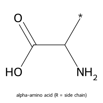
Glycine is the simplest: its R group is just , making it the only amino acid with no chiral center (two H's off the α-carbon). Common MCAT trick.
Quick check: Why does glycine lack optical isomers?
Answer: The α-carbon of glycine bears two identical hydrogen substituents, so it is not a stereocenter — no enantiomers exist.
Absolute Configuration at the α-Carbon
Must knowAll 20 proteinogenic amino acids are L — ribosomes incorporate only the L-form. In a Fischer projection (carboxyl on top, R on bottom), L has the on the left, D on the right. (D-amino acids appear in some bacterial cell walls but not human proteins.)
The more rigorous R/S (CIP) designation tracks the same molecules: nearly all L-amino acids are (S). The one exception to know: L-cysteine is (R), because its side chain has a sulfur that outranks the carboxyl in CIP priority, flipping the label even though the spatial arrangement is the same L-type. You do not need to work CIP priorities for each amino acid.
Quick check: A student says "L-cysteine must be (S) because all L-amino acids are (S)." What is wrong with this statement?
Answer: L-cysteine is (R) because the sulfur-containing side chain ranks higher in CIP priority than the carboxyl group, reversing the R/S designation even though the physical arrangement is the same L-type.
Amino Acids as Dipolar Ions (Zwitterions)
Why Amino Acids Exist as Zwitterions
Must knowNear physiological pH the amino group is protonated (base) and the carboxyl is deprotonated (acid) simultaneously, giving a zwitterion — both a + and − charge but zero net charge. This is the dominant form at pH ~7.4 for simple amino acids.
pKa Values
Must know(ranges only — exact decimals not required)
| Group | Typical pKa |
|---|---|
| -Carboxyl () | ~2 |
| -Amino () | ~9–10 |
| Acidic R groups (Asp, Glu) | ~4 |
| Basic R groups (Lys, Arg, His) | ~6–12.5 |
Isoelectric Point (pI)
Must knowThe isoelectric point is the pH at which the amino acid carries zero net charge (exists as the zwitterion); at the pI it does not migrate in an electric field and has minimal solubility — the basis for isoelectric focusing.
The rule: average the two pKa values that flank the neutral form.
- No ionizable side chain: (the α-COOH and α-NH₃⁺).
- Acidic (Asp, Glu): average the two carboxyl pKas → pI is low (acidic).
- Basic (Lys, Arg, His): average the two amino pKas → pI is high (basic).
Worked Example — pI of aspartic acid (pKa values 1.9, 3.7, 9.6). At net-zero charge, the α-amino is +1, the more-acidic α-carboxyl is −1, and the side-chain carboxyl stays protonated (0). The zwitterion is flanked by and , so:
Asp is acidic, so pI < 7 ✓ — it is negatively charged at physiological pH.
Quick check: Would lysine's pI be above or below 7? Why?
Answer: Above 7. Lysine has a basic side chain (, pKa ~10.5). Its pI is the average of the two amino group pKas (~9.0 and ~10.5), giving ~9.7. You need a high pH to neutralize both positive charges.
Reading the Titration Curve
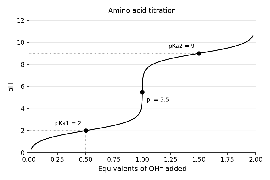
Know the logicA titration curve plots pH vs. equivalents of base added. Each ionizable group gives a buffering plateau centered on its , where the group is half-protonated () and buffering is maximal. Between plateaus the curve rises steeply through an equivalence point. The pI is the steep midpoint between the two plateaus flanking the zwitterion (the average of those two pKas). A simple amino acid shows two plateaus; one with an ionizable side chain shows three. You should be able to read each , locate equivalence points, and find net charge at any pH from such a plot.
Classification of the 20 Amino Acids
Must knowthe 20 amino acids, their one/three-letter codes, and side-chain character. Organized by side chain below.
Nonpolar / Hydrophobic
Must knowAliphatic or aromatic hydrocarbon side chains; cluster in the protein interior.
| Name | 3-Letter | 1-Letter | Side Chain Notes |
|---|---|---|---|
| Glycine | Gly | G | ; achiral; smallest |
| Alanine | Ala | A | |
| Valine | Val | V | branched |
| Leucine | Leu | L | branched |
| Isoleucine | Ile | I | branched |
| Methionine | Met | M | thioether (), not thiol; start codon |
| Proline | Pro | P | cyclic, secondary amine; helix breaker |
| Phenylalanine | Phe | F | benzyl |
| Tryptophan | Trp | W | indole; largest; absorbs 280 nm |
Two special-behavior residues: Proline cyclizes onto the backbone N, rigidly kinking the chain and breaking α-helices (often the first residue of a β-turn). Glycine (R = ) has the least steric hindrance and maximal backbone flexibility, so it sits at tight turns and hinges. Proline constrains; glycine loosens.
Polar, Uncharged
Must knowH-bond with water but carry no net charge near pH 7.
| Name | 3-Letter | 1-Letter | Side Chain Notes |
|---|---|---|---|
| Serine | Ser | S | ; phosphorylation site |
| Threonine | Thr | T | ; phosphorylation site |
| Cysteine | Cys | C | ; thiol; disulfide bonds |
| Tyrosine | Tyr | Y | phenol; phosphorylation site; absorbs 280 nm |
| Asparagine | Asn | N | amide of aspartate |
| Glutamine | Gln | Q | amide of glutamate |
Acidic (Negative at pH 7)
Must knowCarboxyl side chains, deprotonated at physiological pH → net −1.
| Name | 3-Letter | 1-Letter | R-group pKa |
|---|---|---|---|
| Aspartate | Asp | D | ~4 |
| Glutamate | Glu | E | ~4 |
Basic (Positive at pH 7)
Must know| Name | 3-Letter | 1-Letter | R-group pKa | Notes |
|---|---|---|---|---|
| Lysine | Lys | K | ~10.5 | long amine chain |
| Arginine | Arg | R | ~12.5 | most basic; always + at physiological pH |
| Histidine | His | H | ~6.0 | imidazole; only pKa near physiological pH |
Histidine is MCAT-beloved: its side-chain pKa (~6) sits near physiological pH, so it flips between charged and neutral with small pH shifts — the ideal proton shuttle in enzyme active sites (Bohr effect, serine proteases).
Essential vs. Nonessential
Passage-levelEssential amino acids must come from the diet; nonessential are made endogenously. The nine essential: Phe, Val, Thr, Trp, Ile, Met, His, Leu, Lys (mnemonic "PVT TIM HALL"). Arg and His are conditionally essential (needed from diet during growth/illness).
Aromatic
Must knowPhe, Tyr, Trp are aromatic. Trp and Tyr absorb strongly at 280 nm — the basis for measuring protein concentration (A₂₈₀); Phe absorbs weakly.
Quick check: An enzyme's active site contains a residue that acts as a proton donor at pH 6.5 but neutral at pH 8. Which amino acid is most likely responsible?
Answer: Histidine. Its imidazole side chain has pKa ~6.0, so it is ~protonated (charged, proton donor) below 6 and neutral above ~7. It is the only standard amino acid with a pKa near physiological range.
Amino Acids as Hydrophobic or Hydrophilic
Must knowThe hydrophobic/hydrophilic split predicts folding: hydrophobic residues bury in the core to escape water (hydrophobic effect); hydrophilic residues face the aqueous surface.
- Hydrophobic: Gly, Ala, Val, Leu, Ile, Pro, Phe, Met, Trp
- Hydrophilic: all charged residues + polar uncharged (Ser, Thr, Cys, Tyr, Asn, Gln)
(Cys is occasionally listed as weakly hydrophobic, but the MCAT generally treats it as polar.)
Quick check: You mutate a buried valine to glutamate in a globular protein. Predict the effect on stability.
Answer: Likely destabilizing. Glutamate is charged/hydrophilic and disfavors burial in the hydrophobic core. Placing a negatively charged residue away from water is energetically costly, likely unfolding or disrupting local structure.
Key Reactions of Amino Acids
Sulfur Linkage: Cysteine and Cystine
Must knowCysteine's thiol () is the most reactive side chain. Two cysteines oxidize to a disulfide bond (), forming cystine; reduction reverses it.
- Disulfides are covalent, stabilizing tertiary/quaternary structure (intramolecular within a chain; intermolecular linking chains, e.g., insulin's A/B chains).
- They form in the oxidizing ER, not the reducing cytoplasm — so secreted/membrane proteins have them, cytoplasmic ones generally don't.
- Reducing agents (DTT, β-mercaptoethanol; glutathione in vivo) break them.
Quick check: When a researcher treats a disulfide-bonded protein with excess β-mercaptoethanol, what happens?
Answer: The disulfide bonds are reduced to free thiols. The protein loses its disulfide-mediated cross-links, often leading to unfolding/denaturation. This is the principle behind SDS-PAGE when run under reducing conditions.
Peptide Bond Formation and Polypeptides
Must knowA peptide bond () forms when one amino acid's α-amino reacts with another's α-carboxyl, losing water — a condensation (dehydration) reaction.
Know the logic
- Partial double-bond character from resonance (carbonyl O–C–N), making the bond planar with restricted rotation about C–N.
- Predominantly trans (the two α-carbons on opposite sides), minimizing steric clash. Optional cis appears at some Xaa–Pro bonds.
- Rotation is allowed about the (φ) and (ψ) bonds — the degrees of freedom defining secondary structure (Ramachandran plot).
Polypeptide conventions:
- N-terminus (free ) written left; C-terminus (free ) written right; numbered from the N-terminus.
- An n-residue peptide has (n − 1) peptide bonds and releases (n − 1) waters.
Quick check: A tetrapeptide (4 amino acids) is synthesized on a ribosome. How many peptide bonds does it contain, and at what pH would you expect the molecule to carry net zero charge if all four residues have non-ionizable side chains?
Answer: 3 peptide bonds. With non-ionizable side chains, only the terminal -amino (pKa ~9) and -carboxyl (pKa ~3) matter; the internal backbone amides/carbonyls are not ionizable. The pI ≈ (3 + 9)/2 = 6.
Hydrolysis of the Peptide Bond
Must knowPeptide bonds are thermodynamically unstable but kinetically stable (resonance/planarity create a high barrier), so they persist without a catalyst. Hydrolysis adds water back across the bond:
Passage-level
Ways to cleave: strong acid (6 M HCl, destroys Trp); base (causes racemization); or proteases, which are sequence-specific — trypsin cuts C-terminal to Lys/Arg (basic), chymotrypsin cuts after large aromatics (Phe/Tyr/Trp). Each cleavage consumes one water.
Quick check: A researcher performs a complete acid hydrolysis of a decapeptide (10 residues). How many water molecules are consumed?
Answer: 9. There are 9 peptide bonds, each requiring one water for hydrolysis.
Common Confusions & Tricks
1. "All L-amino acids are (S)" — FALSE. L-Cysteine is (R) because sulfur outranks oxygen in CIP priority. Every other standard L-amino acid is (S) at the α-carbon. Cysteine is the one to know by name.
2. Zwitterion vs. neutral molecule — The fully neutral form () barely exists; in solution amino acids are zwitterions with the proton transferred internally. Net charge is zero at the pI, but both charges are present. Don't draw the "neutral" form.
3. Acidic amino acids have LOW pI; basic have HIGH pI — Extra acid groups (Asp, Glu) need more acid (lower pH) to reach zero charge → low pI (~3). Extra amine groups (Lys, Arg) need more base → high pI (~9–11).
4. Asparagine ≠ Aspartate; Glutamine ≠ Glutamate — Asp/Glu have ionizable acidic carboxyl side chains; Asn/Gln are their neutral amides. Trick: N and Q have No charge; D and E are charged.
5. Disulfide bonds ≠ strongest interaction — Covalent and strong, but only relevant to tertiary/quaternary structure; don't confuse with the backbone peptide bond.
6. Methionine vs. Cysteine sulfur — Met has a thioether () and cannot form disulfides; Cys has a thiol () and can. Met is the start codon amino acid (AUG).
7. Proline in helices — Proline breaks α-helices: its ring nitrogen has no N–H to donate to the helix H-bond and it rigidly kinks the backbone. A proline mutation mid-helix → predict disruption.
8. Histidine is the MCAT's favorite — Bohr effect, serine protease catalytic triad (Ser-His-Asp), buffering near physiological pH, zinc coordination. pH-dependent enzyme activity near pH 6–7 → think His.
9. Henderson-Hasselbalch for protonation state — If , the protonated form dominates; if , the deprotonated form dominates. Amino: protonated = ; carboxyl: protonated = .
10. Peptide bond is drawn C→N — From one residue's carbonyl carbon to the next residue's nitrogen. The amide nitrogen is not basic — its lone pair is delocalized into the carbonyl.
Key Equations
| Equation | Variables & When to Use |
|---|---|
| Average the two pKas that flank the zwitterion. Simple: α-COOH and α-NH₃⁺. Acidic: two carboxyls. Basic: two amines. | |
| Henderson-Hasselbalch: ratio of deprotonated to protonated form at a given pH → predominant charge state of any group. | |
| Oxidative disulfide formation (cysteine → cystine); reversed by reducing agents. | |
| Hydrolysis of one peptide bond; consumes 1 water. | |
| Peptide bonds in -residue chain | Also the number of waters released in synthesis. |