What Enzymes Are and Why They Matter
Must knowPriority labels: Must know = cold; Know the logic = mechanism not names; Passage-level = recognize, don't memorize; Optional = skippable.
An enzyme is a biological catalyst, almost always a protein, that accelerates a specific chemical reaction without being consumed by it. Most enzymes are proteins, but some catalytic RNA molecules — ribozymes (e.g., the ribosome's peptidyl transferase activity) — are catalysts too. The defining feature is catalysis, not chemical identity.
An enzyme cannot make an unfavorable reaction favorable. It does not change the overall free energy difference () between reactants and products; it changes only how quickly the reaction reaches equilibrium, by lowering the energy barrier. The MCAT tests this repeatedly. Because every metabolic pathway (glycolysis, the TCA cycle, etc.) is a cascade of enzyme-catalyzed steps, reactions that take milliseconds with enzymes would take centuries without them.
Quick check: An enzyme that is fully saturated with substrate produces product at a maximum rate. You then add more enzyme to the reaction. What happens to the reaction rate?
Answer: The overall rate increases, because now there are more enzyme molecules available to catalyze the reaction, even though each individual enzyme was already working as fast as it could. Adding more enzyme effectively increases the total "ceiling."
Enzyme Classification by Reaction Type
Must knowBuild a mental model of the chemical "job" each does; don't memorize EC numbers.
| Class | What it does | Classic example |
|---|---|---|
| Oxidoreductases | Transfer electrons (oxidation/reduction) | Lactate dehydrogenase, alcohol dehydrogenase |
| Transferases | Move a functional group from one molecule to another | Kinases (transfer phosphate), transaminases |
| Hydrolases | Break bonds using water | Proteases, lipases, phosphatases |
| Lyases | Break bonds without water or oxidation (or form bonds by reverse) | Pyruvate decarboxylase, aldolase |
| Isomerases | Interconvert structural or geometric isomers | Triose phosphate isomerase, mutases |
| Ligases | Join two molecules, requiring ATP hydrolysis | DNA ligase, aminoacyl-tRNA synthetases |
High-yield connections: kinases (transferases) add a phosphate from ATP; phosphatases (hydrolases) remove it by hydrolysis — an antagonistic pair central to signaling. Proteases are hydrolases that cleave peptide bonds (digestion: pepsin, trypsin, chymotrypsin).
Quick check: Aldolase cleaves fructose-1,6-bisphosphate into two three-carbon fragments during glycolysis — no water or redox chemistry involved. What enzyme class does aldolase belong to?
Answer: Lyase. It breaks a C–C bond without water and without oxidation/reduction.
Reduction of Activation Energy
Must knowEvery reaction has a "hill" separating reactants from products. The top is the transition state — the fleeting, high-energy arrangement the reaction must pass through; the height from the reactant side is the activation energy ().
Enzymes lower by stabilizing the transition state. The enzyme binds the transition state more tightly than it binds substrate or product, lowering the barrier so more molecules can cross it per unit time — this differential binding is the thermodynamic basis of catalysis. (This is why transition-state analogs make potent inhibitors: the enzyme binds them tightly but cannot complete the reaction.)
The reaction-coordinate diagram below makes the key point visual: the enzyme lowers the barrier () but leaves the reactant and product free-energy levels — and therefore the overall — unchanged.
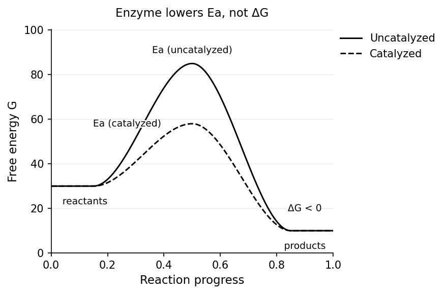
The of the overall reaction (the difference between the final and initial free energy levels) is identical whether the enzyme is present or not.
Quick check: A poison permanently destroys an enzyme in a pathway. The overall for that pathway's reaction is −25 kJ/mol. After the enzyme is destroyed, what is the for the same reaction?
Answer: Still −25 kJ/mol. Enzymes affect rate (kinetics), not the thermodynamic favorability () of the reaction.
Substrates and Enzyme Specificity
Must knowThe molecule an enzyme acts on is its substrate. Specificity — the selective relationship between enzyme and substrate — arises entirely from the enzyme's 3-D structure (set by primary sequence, maintained by higher-order folding). Denaturing an enzyme destroys its shape and its activity even though primary-sequence bonds stay intact.
Specificity spans a range: absolute (one substrate only, e.g., urease), group (a shared functional group, e.g., hexokinase on several hexoses), and stereochemical. Because the active site is a chiral environment built from L-amino acids, enzymes distinguish enantiomers — e.g., amino-acid-metabolizing enzymes act on L- but not D-amino acids.
Quick check: A researcher synthesizes an enzyme's substrate as a pure D-enantiomer instead of the natural L-form. What do you predict will happen when this D-substrate is mixed with the enzyme?
Answer: Little to no catalysis. The enzyme's active site has a specific three-dimensional shape that complements only the correct enantiomer. The D-form cannot form the proper contacts needed for binding and catalysis.
Active Site Model (Lock and Key)
Must knowThe active site is the pocket where substrate binds and catalysis occurs — a cleft formed by a small subset of residues brought together by the tertiary fold; the rest of the protein mostly maintains that geometry.
The active site is a rigid lock perfectly complementary to the substrate "key," with geometry pre-optimized before binding. It explains specificity but wrongly assumes a static, pre-formed site — the limitation the induced-fit model corrects.
Quick check: The lock-and-key model predicts that the enzyme's active site shape is complementary to the substrate. But catalysis requires preferential stabilization of the transition state. Does the lock-and-key model adequately explain transition-state stabilization?
Answer: Not really. If the active site is perfectly complementary to the substrate's ground-state geometry (not the transition state), the enzyme would bind substrate and product equally well but would not specifically stabilize the transition state. This is one of the conceptual weaknesses of the rigid lock-and-key picture.
Induced-Fit Model
Must knowThe active site is not perfectly pre-formed; substrate binding induces a conformational change that aligns catalytic residues, and both molecules adjust to optimize catalytic geometry. This is the more accurate model. It explains transition-state stabilization better (the enzyme moves toward a conformation that stabilizes the transition state, not just the ground-state substrate).
Passage-levelInduced fit also underlies allosteric regulation, where an effector binds a site distant from the active site and reshapes it. A key example is feedback inhibition, where a pathway's end product inhibits the pathway's first enzyme. On test day, choose induced fit whenever evidence shows the enzyme changes shape on binding.
Quick check: Hexokinase (which phosphorylates glucose) is known to close around its substrate upon binding, dramatically changing its shape and excluding water from the active site. Which model does this support, and why does excluding water matter?
Answer: Induced-fit model. The closure is a conformational change triggered by substrate binding. Excluding water prevents ATP hydrolysis (which would be wasteful) and ensures the phosphoryl group is transferred to glucose rather than to water.
Mechanism of Catalysis
Know the logicKnow these strategies qualitatively (what each does); a single enzyme often combines several. Don't memorize residue-level mechanisms.
- Acid–base catalysis: side chains donate/accept protons to facilitate bond breaking/forming. Histidine (pKa ≈ 6, near physiological pH) is the classic general acid-base residue.
- Covalent catalysis: the enzyme forms a transient covalent bond with the substrate, making a more reactive intermediate that is then resolved, regenerating the enzyme.
- Metal ion catalysis: an active-site metal stabilizes charge, acts as an electrophile, or enables redox. Example: carbonic anhydrase uses to catalyze (central to blood CO₂ transport — a favorite MCAT integration).
- Proximity/orientation: holding substrates together in the right alignment raises their effective concentration and orbital overlap.
- Electrostatic/microenvironment: the active site offers a tailored (often less polar) environment that stabilizes charged transition states and shifts residue pKa values.
Quick check: In chymotrypsin's catalytic mechanism, the serine residue forms a covalent bond with the substrate. After the reaction is complete, is the enzyme the same as it was before? Does this violate the definition of a catalyst?
Answer: Yes, the enzyme is regenerated — the serine is eventually freed from the acyl-enzyme intermediate via hydrolysis, restoring the active site to its original state. This is consistent with the definition of a catalyst: it participates in the mechanism but is not net consumed.
Cofactors and Coenzymes
Must knowMany enzymes need a non-protein helper to be active. holoenzyme = active enzyme + cofactor; apoenzyme = protein alone (inactive); a cofactor bound tightly/permanently is a prosthetic group.
- Cofactors are inorganic ions (e.g., , , ). They often help catalysis directly — e.g., Mg²⁺ neutralizes ATP's phosphate charge, making the terminal phosphate a better electrophile for transfer.
- Coenzymes are organic helpers, often vitamin-derived, that carry chemical groups or electrons between steps. Some bind tightly (prosthetic groups, e.g., heme); others bind loosely and cycle as a "co-substrate" (e.g., NAD⁺).
Recognize the major carriers and what they shuttle; don't memorize every structure: NAD⁺/NADH and FAD/FADH₂ (electron carriers), NADPH (electron carrier for anabolism), CoA (acyl groups, e.g., acetyl-CoA), TPP (decarboxylation of α-keto acids), PLP (amino-group transfer), THF (one-carbon transfers), B12 (methyl transfer), biotin (CO₂ carrier in carboxylations).
Quick check: Pyruvate dehydrogenase requires several coenzymes. One facilitates oxidative decarboxylation of an α-keto acid. Which coenzyme is responsible for the decarboxylation step?
Answer: Thiamine pyrophosphate (TPP), which facilitates decarboxylation of α-keto acids. (PDH's other coenzymes — CoA, FAD, NAD⁺, and lipoic acid — handle acyl transfer and electron flow.)
Water-Soluble Vitamins
Must knowMost coenzymes derive from vitamins, so the MCAT links enzyme function to vitamin biology. Water-soluble vitamins (B vitamins, vitamin C) aren't stored in quantity and must be eaten regularly; deficiency impairs the enzymatic step that depends on them. You don't need to memorize the deficiency-syndrome table — focus on the two high-yield items below.
Vitamin C (ascorbic acid) acts as a reducing agent and is required by prolyl hydroxylase to hydroxylate proline/lysine in collagen synthesis. Deficiency → scurvy (weak connective tissue, bleeding gums, poor wound healing).
Folate vs. B12: both deficiencies cause megaloblastic anemia, but B12 deficiency additionally causes neurological damage (spinal-cord demyelination) and elevated methylmalonic acid (MMA), because B12 is needed by methylmalonyl-CoA mutase. Folate deficiency does neither.
Quick check: A patient has megaloblastic anemia but no neurological symptoms, and serum methylmalonic acid (MMA) is normal. Folate or B12 deficiency?
Answer: Folate. B12 deficiency would raise MMA and produce neurological symptoms; folate deficiency disrupts one-carbon metabolism without affecting the MMA pathway or myelination.
Effects of Local Conditions on Enzyme Activity
Must knowEnzyme activity is sensitive to the local environment. The major variables on MCAT passages:
Temperature. Rate rises with temperature (more productive collisions) up to an optimum (~37°C for human enzymes). Above it the enzyme denatures — non-covalent interactions break, active-site geometry is lost, activity plummets (why high fever is dangerous).
pH. Each enzyme has an optimal pH; off-optimum pH changes the protonation of active-site side chains (disrupting binding/catalysis), and extremes can denature. Classic examples: pepsin ~2 (acidic stomach); trypsin/chymotrypsin ~8 (alkaline small intestine); salivary amylase ~7.
Substrate concentration. At fixed enzyme, rate rises with — nearly linearly at first, then leveling off as active sites fill until the enzyme is saturated at . This gives the hyperbolic rate-vs- curve, parameterized by (half-maximal ; inverse measure of affinity) and .
The quantitative kinetics framework (Michaelis–Menten, Lineweaver–Burk, cooperativity, inhibitor types) is developed in the Control of Enzyme Activity guide. Here, just know that substrate/cofactor availability is one of the local conditions setting enzyme rate.
Common Confusions & Tricks
1. Enzymes don't change — only . If a question says "the enzyme makes the reaction thermodynamically favorable," that is false. Enzymes cannot change , , or the direction of spontaneity. They only speed up equilibration.
2. Enzymes lower by stabilizing the transition state, not the substrate. A perfectly substrate-complementary site would bind substrate and product equally and give no catalytic boost. Catalysis comes from binding the transition state more tightly than either — which is why transition-state analogs are such potent inhibitors.
3. Apoenzyme vs. holoenzyme. Apo = "without" (apoenzyme lacks its cofactor/coenzyme), Holo = "whole" (holoenzyme has everything it needs). The apo- prefix means "away from" — same root as "apoptosis" (cell falling away).
4. Coenzymes are NOT consumed. They are regenerated elsewhere in metabolism. NAD⁺ is reduced to NADH, then reoxidized by the electron transport chain. They cycle; they are not net consumed.
5. Cofactor (inorganic) vs. coenzyme (organic). A cofactor is an inorganic ion (Zn²⁺, Mg²⁺, Fe²⁺); a coenzyme is an organic helper molecule, often vitamin-derived (NAD⁺ from niacin, FAD from riboflavin, TPP from thiamine). Both restore activity to an otherwise inactive apoenzyme.
6. Lock-and-key says active site is complementary to the substrate; induced fit says the enzyme changes shape upon binding. If a passage describes an enzyme changing conformation, think induced fit. If it emphasizes rigid geometric complementarity, think lock-and-key.
7. Temperature and pH each have an optimum. Past the optimum, rate falls because the enzyme denatures (loses the active-site geometry), not because the chemistry reverses. Extreme pH also changes the ionization of catalytic side chains.
Key Equations
| Equation | Variables & when to use |
|---|---|
| Enzyme mechanism: enzyme binds substrate to form complex , then releases product and regenerates | |
| Enzymes lower activation energy () without changing |
(The quantitative kinetics equations — Michaelis–Menten, Lineweaver–Burk, , and the Hill equation — are listed in the Control of Enzyme Activity guide.)