Proteins do far more than catalyze reactions. Most proteins never act as enzymes — they bind things, defend against threats, and physically move cargo. Think of them as molecular machines shaped by their structure to do a mechanical, communicative, or structural job. This guide covers the three MCAT-tested domains: binding/protein–ligand interactions, immune system proteins, and motor proteins.
Priority labels: Must know = cold; Know the logic = mechanism not names; Passage-level = recognize, don't memorize; Optional = skippable.
Binding
The Core Idea: Shape as Function
Must knowA binding protein has a specific three-dimensional binding site — a pocket shaped by its tertiary (and sometimes quaternary) structure to be complementary to a particular ligand (any molecule that binds noncovalently). The fit is flexible and dynamic (induced-fit model), not a rigid lock-and-key, but specificity is real.
Know the logicBinding affinity is measured by the dissociation constant (units of concentration). A lower means higher affinity — the complex falls apart less readily. Fractional saturation (, the fraction of sites occupied) traces a hyperbolic binding curve for simple binding, and by definition the sites are half-occupied when . A lower- protein reaches high saturation at much lower ligand concentration. (The MCAT tests this comparison qualitatively; explicit calculations are out of scope.)
Hemoglobin vs. Myoglobin: The Canonical Binding Example
Must knowThe single most-tested binding example is hemoglobin (Hb) vs. myoglobin (Mb).
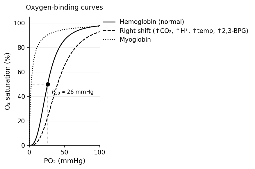
Myoglobin is monomeric (one heme), binds with a hyperbolic curve and very high affinity (low ), and stores oxygen in muscle.
Hemoglobin is a tetramer ( in adults) and binds with a sigmoidal (S-shaped) curve — the hallmark of cooperative binding: binding of one raises the affinity of the remaining subunits. Mechanistically, Hb shifts between a low-affinity T-state (tense, deoxy) and a high-affinity R-state (relaxed, oxy); each bound pushes the tetramer toward R, easing the next binding (homotropic allosteric regulation). The degree of cooperativity is captured by the Hill coefficient ( = none/hyperbolic; = positive/sigmoidal, Hb ).
Cooperativity lets Hb act like a switch: it loads efficiently in the lungs (high ) and unloads it in tissues (low ). On a curve, Mb's hyperbola sits far left (high affinity); Hb's sigmoid lies right and shifts further right under Bohr conditions.
The Bohr Effect and Allosteric Modulators of Hemoglobin
Must knowSeveral heterotropic allosteric effectors shift Hb's curve without being the transported ligand. All four below lower Hb's affinity (shift right), promoting unloading in active tissues; each stabilizes the T-state:
- (forms carbaminohemoglobin) — active tissues make
- pH / H⁺ (the Bohr effect) — active tissues make lactic acid and
- temperature — warm exercising muscle
- 2,3-BPG (binds deoxy-Hb central cavity) — altitude, chronic anemia
So in the lungs (high pH, low ) the curve shifts left and Hb loads ; in tissues (low pH, high ) it shifts right and unloads — self-regulating.
Carbon monoxide () binds heme far more tightly than AND left-shifts the remaining subunits (locks them in R), so they won't release their — the double hit that makes CO poisoning deadly.
Fetal hemoglobin (HbF, ) is a high-yield application: its subunits bind 2,3-BPG weakly, giving HbF higher affinity (left-shifted, lower P₅₀) than adult HbA — so the fetus pulls across the placenta down the affinity gradient.
Other Transport and Storage Proteins
Must knowAlbumin is the most abundant plasma protein and the body's general-purpose blood carrier (fatty acids, bilirubin, steroid hormones, many drugs); it is also the main contributor to plasma oncotic (colloid osmotic) pressure. For ligands carried on albumin, only the free (unbound) fraction is biologically active — a recurring pharmacology theme.
Receptors
Must knowA receptor binds a specific signaling ligand (hormone, neurotransmitter) and transduces that event into a cellular signal. Cell-surface receptors (GPCRs, receptor tyrosine kinases, ligand-gated ion channels) bind hydrophilic ligands that can't cross the membrane and relay the signal inward by conformational change. Intracellular receptors bind small hydrophobic ligands (e.g., steroid hormones) that diffuse through the membrane, and the complex often acts directly as a transcription factor. The same binding logic (specificity, affinity) applies throughout.
Structural Proteins as Binding/Scaffolding Proteins
Must knowStructural proteins bind each other and cells to provide mechanical support.
- Collagen — most abundant protein in the body; a triple helix giving tensile strength to bone, tendon, skin, cartilage. Its synthesis needs vitamin C (for hydroxyproline) — deficiency causes scurvy.
- Keratin — α-helical fibrous protein of skin, hair, and nails; strength comes partly from disulfide bonds (cysteine-rich).
- Passage-level elastin (stretch/recoil in arteries, lung, skin); fibronectin and laminin (ECM adhesion proteins linking cells to matrix via integrins).
Quick check: Myoglobin has a for of approximately 1 mmHg (in terms of partial pressure), while hemoglobin's P₅₀ (the at 50% saturation) is about 26 mmHg. Which protein is more suitable for storing oxygen in muscle, and why?
Answer: Myoglobin. Its higher affinity (lower ) keeps it nearly saturated even at the low of resting muscle, while hemoglobin readily releases to it at tissue oxygen tensions — a coordinated handoff.
Immune System
Organizing the Immune Response
Must knowImmunity has two arms:
- Innate — fast, nonspecific, no memory: barriers, phagocytes (macrophages, neutrophils), NK cells, inflammation, and the complement system.
- Adaptive — slower but specific, with memory. It splits into humoral immunity (B cells + secreted antibodies; targets pathogens in fluids) and cell-mediated immunity (T cells; targets infected/abnormal cells).
Both B and T cells arise in bone marrow, but B cells mature in the Bone marrow and T cells mature in the Thymus. Helper T cells (CD4⁺) coordinate via cytokines; cytotoxic T cells (CD8⁺) kill infected/abnormal cells; regulatory T cells dampen the response.
Antibody Structure: A Protein Designed for Binding
Must knowThe MCAT focuses on antibodies (immunoglobulins) and, secondarily, MHC. An antibody is a Y-shaped glycoprotein of four chains — two heavy (H) and two light (L) — joined by disulfide bonds. Each chain has a constant (C) region (the Y's stem; same within a class; sets effector function) and a variable (V) region (the tips; unique per clone; forms the antigen-binding site).
Two functional regions:
- Fab (antigen-binding): the two arms; each antibody has two identical binding sites (bivalent). The pocket is formed by hypervariable loops (CDRs) in the V regions.
- Fc (crystallizable): the constant stem; binds Fc receptors on immune cells and triggers complement, opsonization, and ADCC.
Antigen–antibody binding is noncovalent but collectively tight. The antigen region actually bound is the epitope; the matching antibody site is the paratope.
Antibody binding produces several outcomes: neutralization (coating a toxin/virus to block host entry), agglutination (cross-linking cells into clumps), precipitation (cross-linking soluble antigens into aggregates), opsonization (coating a pathogen to flag it for phagocytosis), and complement activation.
The Complement System
Passage-levelComplement is a plasma-protein cascade (innate) that antibodies can trigger. Know its three outcomes: opsonization, inflammation, and lysis via the membrane attack complex. (The detailed cascade biochemistry is out of scope.)
The Five Immunoglobulin Classes
Know the logicThe five isotypes differ in their heavy-chain constant regions:
| Isotype | Location / Form | Key Function |
|---|---|---|
| IgG | Most abundant in serum; monomer | Opsonization, complement, crosses placenta (passive immunity to fetus) |
| IgM | Pentamer (10 antigen-binding sites) | First antibody produced in primary response; efficient complement activation |
| IgA | Dimer in secretions (saliva, breast milk, mucosa) | Mucosal immunity; passive immunity via breast milk |
| IgE | Monomer, very low serum concentration | Binds mast cells/basophils; allergy and anti-parasitic responses |
| IgD | Monomer on naïve B-cell surface | B-cell receptor; role in B-cell activation (less tested) |
A mnemonic: GAMED (G, A, M, E, D) or remember the abundance order: IgG > IgA > IgM > IgE > IgD.
Clonal Selection and Immunological Memory
Must knowEach B cell expresses one unique antibody specificity (clonal selection). When antigen binds its surface receptor and a T helper (CD4⁺) cell helps, the B cell undergoes clonal expansion into plasma cells (antibody-secreting factories) and long-lived memory B cells. This drives the difference between the primary response (slow, lower titer, IgM first) and the secondary response (faster, higher titer, mostly IgG, from memory cells) — the basis of how vaccines work. (Class switching keeps the same specificity while swapping the heavy-chain constant region; the recombination mechanism is out of scope.)
MHC Molecules: Presenting Antigens to T Cells
Must knowMHC molecules are cell-surface glycoproteins that display peptide fragments for T-cell surveillance — how T cells "see" inside cells.
- MHC class I: on all nucleated cells; displays intracellular (incl. viral) peptides; recognized by cytotoxic T cells (CD8⁺) → infected cell killed.
- MHC class II: only on professional APCs (dendritic cells, macrophages, B cells); displays internalized extracellular peptides; recognized by helper T cells (CD4⁺) → cytokine secretion, B-cell/macrophage activation.
Mnemonic: MHC I → cells that kIll (CD8); MHC II → Helpers (CD4).
Passage-levelMHC underlies self/non-self discrimination: thymic training establishes self-tolerance (self-reactive T cells deleted); its breakdown causes autoimmunity. In transplantation, donor MHC (HLA) differs from the recipient's, so recipient T cells trigger rejection — hence HLA matching and immunosuppression.
Active vs. Passive Immunity
Must know- Active immunity: The host's immune system responds and generates memory. Can be natural (actual infection) or artificial (vaccination).
- Passive immunity: Pre-formed antibodies are transferred. No memory is generated. Examples: maternal IgG crossing the placenta; IgA in breast milk; therapeutic antibody infusions (e.g., rabies immunoglobulin after exposure). Protection is immediate but temporary.
Quick check: A patient is bitten by a potentially rabid animal and receives both rabies vaccine and rabies immunoglobulin (RIG) simultaneously. Which provides immediate protection, and which will last longer?
Answer: The RIG provides immediate passive protection (pre-formed antibodies against rabies virus). The vaccine stimulates active immunity — it takes days to weeks for antibodies to develop, but the immune memory lasts years.
Motor Proteins in Cellular Movement
The Big Picture: Proteins That Convert Chemistry to Motion
Must knowMotor proteins use ATP hydrolysis to generate directional force, walking along a cytoskeletal track (or moving cargo). Three families:
- Myosin — moves along actin filaments
- Kinesin — moves along microtubules toward the plus end (anterograde, toward periphery)
- Dynein — moves along microtubules toward the minus end (retrograde, toward center); also powers cilia and flagella
Myosin and the Sliding Filament Theory of Muscle Contraction
Must knowMuscle contraction is the most-tested motor topic — know both the sarcomere anatomy and the cross-bridge mechanism.
Sarcomere Structure
Must knowThe sarcomere is the functional unit of skeletal/cardiac muscle, bounded by two Z-discs. Within it:
| Structure | Composition | What happens during contraction |
|---|---|---|
| Thick filaments | Myosin | Stay centered in A-band |
| Thin filaments | Actin + tropomyosin + troponin complex | Slide inward toward M-line |
| A-band | Full length of thick filament | Width stays constant |
| I-band | Thin filament only (no overlap with thick) | Narrows during contraction |
| H-zone | Thick filament only (no overlap with thin) | Narrows during contraction |
| M-line | Center of sarcomere; cross-links thick filaments | Stays at center |
| Z-disc | Anchors thin filaments | Z-discs move closer together |
Critical MCAT fact: the A-band does NOT change width (thick-filament length is constant); the I-band and H-zone shorten and the Z-discs move closer. Filaments slide, they do not shorten — the sliding filament theory.
The Cross-Bridge Cycle
Must knowThe ATP-powered cycle, in four steps:
- ATP binds the myosin head → myosin releases actin (detaches)
- ATP hydrolysis () → head cocks into a high-energy position and weakly rebinds actin
- Power stroke: released → head swings forward, pulling the thin filament toward the M-line; ADP released
- Rigor state: myosin tightly bound with no nucleotide — broken only when new ATP binds
Rigor mortis: after death ATP runs out, so myosin heads can't release actin → muscles lock in the rigor state.
Regulation by Calcium: Troponin and Tropomyosin
Must knowAt rest, tropomyosin blocks the myosin-binding sites on actin. An action potential releases from the sarcoplasmic reticulum (SR):
When the signal stops, the SR pump SERCA (-ATPase) returns to the SR and tropomyosin re-blocks actin. Both contraction and relaxation require ATP.
Kinesin and Dynein: Intracellular Transport Along Microtubules
Must knowMicrotubules are polarized α/β-tubulin polymers: the plus end (fast-growing) points toward the periphery; the minus end is anchored at the centrosome/MTOC.
- Kinesin — plus-end–directed: carries cargo toward the periphery (anterograde).
- Dynein — minus-end–directed: carries cargo toward the cell body (retrograde) and powers cilia/flagella.
In neurons: anterograde (soma → terminal) uses kinesin (e.g., neurotransmitter vesicles); retrograde (terminal → soma) uses dynein.
Cilia and Flagella
Know the logicThe motile axoneme has a "9+2" arrangement (9 doublets + a central pair). Axonemal dynein makes adjacent doublets slide; because nexin links anchor them, they bend rather than slide apart, producing the beat. Primary cilia are non-motile sensory organelles ("9+0," no central pair). Optional defective ciliary motility causes Kartagener syndrome (situs inversus, infertility).
Quick check: In the sliding filament model, a student claims that the A-band shortens during maximal muscle contraction. Are they correct? What does change?
Answer: No. The A-band equals the thick-filament length, which doesn't change. The I-band and H-zone shorten as thin filaments slide inward to overlap the thick filaments, and the sarcomere shortens as the Z-discs are pulled together.
Common Confusions & Tricks
1. Lower = Higher affinity (not lower affinity). Students see a smaller number and think "weaker." Flip your thinking: is a dissociation constant. Smaller → complex stays together → tighter binding. On the MCAT, when comparing two binding proteins, always say "the one with the lower has higher affinity."
2. Hemoglobin's P₅₀ vs. Myoglobin's P₅₀. Hemoglobin has a P₅₀ of ~26 mmHg; myoglobin's is ~1–2 mmHg. Myoglobin is on the left (higher affinity). Students sometimes draw this backward. Remember: Myoglobin is in Muscle, and Muscle stores O₂, so Mb must hold on tightly → leftward, high-affinity curve.
3. Bohr effect shifts the curve RIGHT (promotes O₂ unloading). Low pH, high , high 2,3-BPG, and high temperature all shift right. A right-shift means hemoglobin needs a higher to be 50% saturated → it holds O₂ less tightly → it releases more O₂ to tissues. Never confuse a right-shift with increased affinity.
4. CO poisoning: two mechanisms. CO doesn't just compete with O₂ for the heme — it also causes a left-shift of the remaining three subunits (makes them not release their O₂). Most students only remember the competition part; both effects together are what makes CO so dangerous.
5. A-band does NOT change. In sarcomere questions, the most common trap is "which band shortens?" The A-band stays the same. Only I-band and H-zone shorten. Memorize: A = Always the same.
6. MHC I (CD8) vs. MHC II (CD4). The trick: 1 × 8 = 8 and 2 × 4 = 8 — same product, helps you pair them. Better yet: MHC I presents to cells that kIll (cytotoxic CD8⁺ T cells); MHC II presents to Helper cells (CD4⁺). Also: MHC I is on all nucleated cells (remember this for questions about organ transplant rejection — any nucleated cell can be a target), while MHC II is only on professional APCs.
7. IgM is the first responder; IgG is the memory responder. In a primary response you see IgM rise then fall; in the secondary response IgG dominates. IgG also crosses the placenta (passive immunity in neonates) — IgM cannot, because it is a pentamer (too large).
8. Kinesin = Plus-end = Periphery (anterograde); Dynein = Minus-end = Center (retrograde). In a neuron: kinesin ships cargo OUT (toward terminal); dynein brings cargo BACK (toward soma). For cilia/flagella, dynein is the motor — not kinesin.
9. ATP in muscle: needed for BOTH contraction and relaxation. Rigor mortis is an ATP deficit causing locked contraction. Relaxation requires ATP-powered SERCA to pump back into the SR.
10. Tropomyosin blocks, troponin is the sensor. Students confuse which does what. Tropomyosin physically covers myosin-binding sites on actin. Troponin is the -sensing complex that, when binds troponin C, moves tropomyosin out of the way.
Key Equations
| Equation | Variables and When to Use |
|---|---|
| Dissociation constant; = free protein, = free ligand, = complex. Lower → higher affinity. | |
| Fractional saturation for simple (non-cooperative) binding; when (defines operationally). | |
| Hill equation; = Hill coefficient. → positive cooperativity (sigmoidal). For hemoglobin, . | |
| (cross-bridge cycle) | ATP hydrolysis cocks myosin head; release triggers power stroke; ADP release at end of stroke. Net: 1 ATP per cross-bridge cycle. |
| → tropomyosin shift → actin site exposed | Not a quantitative equation, but the logical sequence for muscle activation; reversed by SERCA (-ATPase) pumping back into SR. |