Proteins are the workhorses of biology — enzymes, structural scaffolds, receptors, transporters, hormones. The MCAT expects you to understand not just what the four levels of structure are, but why each level forms and what forces hold it together. This guide builds from amino acids up to quaternary assemblies, then covers stability, denaturation, and the techniques used to separate proteins.
Priority labels: Must know = cold; Know the logic = mechanism not names; Passage-level = recognize, don't memorize; Optional = skippable.
Amino Acids: The Building Blocks
General Structure
Must knowEvery standard amino acid has a central α-carbon bonded to an amino group (), a carboxyl group (), a hydrogen, and a variable side chain (R group). At physiological pH (~7.4) amino acids are zwitterions — protonated amino group () and deprotonated carboxyl (), net dipolar.
The α-carbon is a chiral center in all amino acids except glycine (R = ). Standard amino acids in proteins are L-amino acids.
The 20 Standard Amino Acids
Know the logicGroup amino acids by side-chain character, not the full table. The categories: nonpolar/hydrophobic (buried in the core), polar uncharged (H-bond, often on the surface), acidic (Asp, Glu — negative at pH 7), and basic (Lys, Arg, His — positive at pH 7). Optional aromatic residues (Phe, Tyr, Trp) absorb UV at 280 nm.
A few side chains carry real testable weight: cysteine forms disulfide bonds; proline's cyclic ring constrains backbone geometry; glycine is the smallest and most flexible; histidine is the only one with a pKa (~6) near physiological pH, making it a common active-site/buffer residue.
Acid-Base Chemistry and pI
Must knowEach amino acid has an α-carboxyl () and α-amino (–10); ionizable R groups add a third pKa.
The isoelectric point (pI) is the pH of zero net charge. For a side-chain-uncharged amino acid:
With an ionizable side chain, average the two pKa values that flank the neutral form: acidic amino acids → the two lowest pKa's; basic amino acids → the two highest.
Worked example — pI of glutamic acid ( = 2.1 α-COOH, 4.1 R-COOH, 9.5 α-amino): the zero-charge form sits between the first two deprotonations, so
Glu is acidic, so pI < 7 — confirmed.
Quick check: At pH 7.4, is glutamic acid positively charged, negatively charged, or neutral?
Answer: Negatively charged. pH 7.4 is well above pI = 3.1, so it has lost protons beyond its isoelectric point and carries net negative charge.
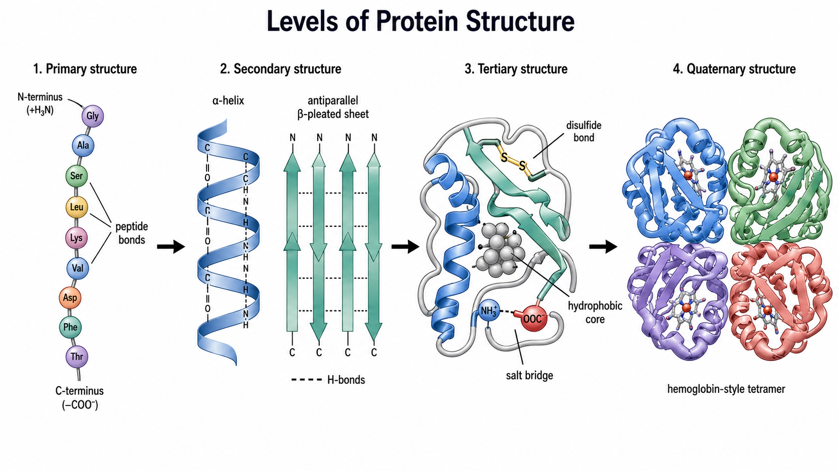
Proteins are organized into four hierarchical levels, each building on the one below. The illustration below summarizes how primary sequence gives rise to local secondary motifs, a folded tertiary shape, and a multi-subunit quaternary assembly.
Primary Structure
Peptide Bond Formation
Must knowAmino acids are joined by peptide bonds, formed via condensation between the carboxyl of one residue and the amino group of the next, releasing water:
A chain of residues is a polypeptide with directionality: free amino end = N-terminus (left by convention), free carboxyl end = C-terminus. A chain of residues has peptide bonds.
Peptide Bond Geometry
Must knowThe peptide bond has partial double-bond character from resonance (carbonyl oxygen ↔ nitrogen lone pair), making it planar and rigid with the trans conformation preferred. Backbone flexibility therefore comes only from rotation around the two single bonds flanking each α-carbon, and only certain combinations are sterically allowed — these give the α-helix and β-sheet.
Sequence as Primary Structure
Must knowPrimary structure is the linear amino acid sequence (N→C), encoded by the gene, that determines all higher-order structure. Same composition but different sequence → entirely different fold. (Experimental sequencing methods are their own topic.)
Quick check: A hexapeptide (6 residues) contains how many peptide bonds?
Answer: peptide bonds.
Secondary Structure
The Concept
Must knowSecondary structure is recurring local backbone geometry stabilized by hydrogen bonds between backbone amide () and carbonyl () groups — the backbone, not the R groups. So it is largely independent of which side chains are present (though proline and bulky residues modulate it).
The α-Helix
Must knowThe α-helix is a right-handed coil in which the carbonyl O of residue H-bonds to the amide NH of residue . There are ~3.6 residues per turn, and R groups project outward while the backbone H-bonds run roughly parallel to the helix axis.
What breaks an α-helix: proline (its ring fixes geometry and removes the backbone NH → "helix breaker," often at turns/hinges), clustered like charges (repulsion), and bulky adjacent R groups.
The β-Sheet
Must knowIn a β-sheet, extended strands align side-by-side with H-bonds between adjacent strands.
- Parallel: strands run the same N→C direction; H-bonds distorted, less stable.
- Antiparallel: strands run opposite directions; H-bonds linear, stronger.
Side chains alternate above and below the sheet plane.
Turns and Loops
Passage-levelβ-turns are short 4-residue reversals (rich in proline and glycine); loops are irregular connecting regions, often surface binding sites.
Quick check: Which type of bond primarily stabilizes secondary structure — disulfide bonds, hydrophobic interactions, or backbone hydrogen bonds?
Answer: Backbone hydrogen bonds. Disulfide bonds and hydrophobic interactions stabilize tertiary structure.
Tertiary Structure
Overview
Must knowTertiary structure is the full 3-D fold of a single polypeptide — how secondary elements pack together — stabilized by side-chain interactions (mostly non-covalent, plus disulfides).
Stabilizing Forces
Must know- Hydrophobic interactions — the dominant driving force. Nonpolar R groups cluster in the interior away from water. This is entropy-driven (expulsion of ordered water), not a direct nonpolar attraction (see stability section).
- Hydrogen bonds — between polar side chains, and side chains and buried water.
- Ionic interactions (salt bridges) — between oppositely charged side chains (e.g., Lys + Asp); prominent on surfaces and disrupted at extreme pH.
- Disulfide bonds () — covalent bonds between the thiols of two cysteine residues via oxidation; the cross-linked pair is cystine. They strongly stabilize structure, especially in secreted/extracellular proteins (oxidizing environment); cytoplasmic cysteines usually stay reduced.
Disulfides are cleaved by reducing agents — β-mercaptoethanol (BME) or DTT — a key fact for denaturation and SDS-PAGE.
Role of proline: its rigid ring restricts backbone rotation, introducing a kink or turn (exploited in collagen and hinge regions).
Fibrous vs. Globular vs. Conjugated Proteins
Must know- Fibrous: long, insoluble, structural, one repetitive secondary motif (e.g., collagen triple helix, keratin, elastin, silk).
- Globular: compact, soluble, hydrophobic core buried (most enzymes, antibodies, hemoglobin).
- Conjugated: carries a non-amino-acid prosthetic group. Optional glycoproteins (carbohydrate), lipoproteins (lipid), metalloproteins (metal), hemoproteins (heme, e.g., hemoglobin).
Quick check
A mutation converts a core cysteine to serine. What stabilizing interaction is most directly lost?
Answer: The disulfide bond (cystine cross-link). Serine has a hydroxyl, not a thiol, so it cannot form bonds.
Quaternary Structure
Definition and Driving Forces
Must knowQuaternary structure is the association of two or more polypeptide subunits into one functional complex, held by the same forces as tertiary structure (hydrophobic, H-bonds, ionic) plus, sometimes, interchain disulfides. Complexes can be homomeric (identical subunits) or heteromeric (e.g., hemoglobin).
Hemoglobin: The Classic Example
Must knowHemoglobin is a heterotetramer (), each subunit carrying a heme group. Cooperative O₂ binding arises from quaternary changes: binding one O₂ shifts the T-state (low-affinity) toward the R-state (high-affinity), easing subsequent binding. Myoglobin (single chain, tertiary only) shows a hyperbolic curve and no cooperativity.
Quick check
A protein has two identical subunits held by hydrophobic contacts and two disulfide bonds between the chains. At which structural level(s) are the disulfide bonds contributing?
Answer: Quaternary — the disulfides are between separate chains (intermolecular), holding subunits together. A disulfide within one chain would contribute to tertiary structure.
Conformational Stability, Denaturing, and Folding
Thermodynamic Basis of Folding
Know the logicFolding is spontaneous under native conditions (), balancing:
- Enthalpy: H-bonds, ionic contacts, and van der Waals in the folded state release energy (favorable).
- Entropy: the chain loses conformational entropy on folding (unfavorable), but this is overcome by the entropy gain of water when hydrophobic groups are buried (next section).
Most proteins are only marginally stable, so modest perturbations can unfold them.
Hydrophobic Interactions and the Solvation Layer
Know the logicWater can't H-bond to a nonpolar surface, so it forms ordered cage-like solvation shells around exposed nonpolar groups, lowering water entropy (unfavorable). Burying those groups in the core releases that ordered water into bulk solvent → large increase in water entropy, which drives folding. So the hydrophobic effect is entropy-driven: it's not nonpolar groups attracting each other, it's liberating ordered water.
Denaturation
Must knowDenaturation is unfolding from the native state. It disrupts 2°/3°/4° structure but does not break peptide bonds, so primary structure is preserved.
Know the logicCommon denaturing agents (not the full table): heat and organic solvents (disrupt H-bonds / hydrophobic effect), urea / guanidinium (compete for H-bonds), pH extremes (disrupt salt bridges and protonation), detergents like SDS (coat with charge, disrupt the core), and reducing agents BME/DTT or heavy metals (attack disulfides). Denaturation can be reversible (refolds on removal) or irreversible (aggregates, like a cooked egg white).
Anfinsen's Experiment — Sequence Determines Structure
Know the logicAnfinsen denatured ribonuclease A with urea + β-mercaptoethanol (unfolding it and cleaving its disulfides), abolishing activity. Slowly removing the denaturants under oxidizing conditions let it spontaneously refold with full activity and correct disulfides. Conclusion: the sequence contains all information for the native fold, and folding is thermodynamically spontaneous (native state = free-energy minimum).
Molecular Chaperones
Passage-levelIn the cell, chaperones (e.g., heat-shock proteins) prevent aggregation of folding polypeptides. They assist folding but do not encode the native structure — the sequence still does.
Quick check: Anfinsen added β-mercaptoethanol to RNase A. What bond type was cleaved, and at which structural level?
Answer: It cleaves disulfide bonds (cystine cross-links) between cysteines, stabilizing tertiary structure (all Cys are within RNase A's single chain).
Separation Techniques
Electrophoresis
Must knowElectrophoresis separates proteins by migration in an electric field. Key variants:
- SDS-PAGE: SDS is an anionic detergent that denatures proteins and coats them with negative charge proportional to size, so separation is by molecular weight only — smaller proteins migrate farther; a MW ladder is run alongside. With BME added, it separates individual chains (reports chain MW, not intact complex MW).
- Native PAGE: no SDS or reducing agent, so proteins keep native charge, shape, and quaternary structure; separates by size + shape + charge. Used when activity must be preserved.
- Isoelectric focusing (IEF): proteins migrate in a pH gradient until they reach their pI (net charge zero), separating by pI.
- 2D gel: IEF (by pI) then SDS-PAGE (by MW) — Passage-level, recognize as a 2-D separation of complex mixtures.
Quick check: A protein has pI 5.0. In SDS-PAGE, does it migrate toward the anode (+) or cathode (−)?
Answer: The anode (+). In SDS-PAGE all proteins are negatively coated by SDS, so all migrate toward the positive electrode regardless of pI.
Isoelectric Point and Electrophoretic Mobility
Must knowIn native electrophoresis: pH > pI → net negative → migrates to the anode (+); pH < pI → net positive → migrates to the cathode (−); pH = pI → no migration. (This is why IEF works.)
Know the logicEstimating net charge: tally every ionizable group — N-terminal amino, C-terminal carboxyl, and each ionizable side chain — using "protonated when pH < pKa, deprotonated when pH > pKa." Protonated amino groups = +1, deprotonated carboxyls = −1; sum them.
Example: Lys-Gly-Asp at pH 7 ≈ N-terminus (+1), Lys (+1), Asp (−1), C-terminus (−1) → net ≈ 0, migrates little; raise pH and it turns net negative.
Chromatographic Separation Techniques
Know the logic- Ion-exchange: a charged resin binds proteins of opposite charge (cation-exchange resin binds + proteins; anion-exchange binds − proteins); eluted by raising salt or changing pH.
- Size-exclusion (gel filtration): porous beads retard small proteins while large ones are excluded and elute first → separates by size; preserves quaternary structure, so it reports native MW.
- Affinity: an immobilized ligand specific to the target binds it while everything else washes through (e.g., His-tag on a nickel column) — the most specific method.
- HPLC: Optional high-pressure column chromatography for high resolution; reversed-phase separates by hydrophobicity.
Quick check: You want to purify a His-tagged protein from a bacterial lysate in one step. Which technique?
Answer: Affinity chromatography (nickel-NTA resin binds the polyhistidine tag; other proteins wash through).
Common Confusions & Tricks
1. Cysteine vs. cystine. Cysteine = one amino acid with a free thiol (). Cystine = two cysteines joined by a disulfide (); named in the context of tertiary stabilization.
2. Secondary-structure H-bonds are backbone, NOT side chains. The α-helix/β-sheet H-bonds are between backbone and . R groups drive tertiary interactions.
3. SDS-PAGE separates by MW, not pI. SDS overrides native charge. Only IEF separates by pI.
4. Proline breaks helices but appears in turns and collagen. It's a helix breaker (no backbone NH, fixed ring) yet abundant in collagen's distinct poly-Pro helix.
5. The hydrophobic effect is entropy-driven. It's not nonpolar attraction — burying nonpolar residues releases ordered solvation-shell water, raising water entropy. Driving force is of water.
6. Disulfide between vs. within chains. Within one chain → tertiary; between different chains → quaternary.
7. Which pKa's to average for pI. Acidic (Asp, Glu): two lowest. Basic (Lys, Arg, His): two highest. Uncharged R: pKa1 and pKa2. (It's the midpoint of the zero-charge plateau.)
8. Anfinsen → sequence determines structure. Spontaneous refolding after denaturation = Anfinsen/thermodynamic hypothesis. Chaperones assist but don't change the sequence's information.
9. Size-exclusion vs. SDS-PAGE for native MW. SDS-PAGE gives individual chain MW; size-exclusion (native) gives intact-complex MW.
10. BME or DTT → disulfide cleavage. These reducing agents cleave bonds; in SDS-PAGE prep (with SDS + heat) they ensure full denaturation.
Key Equations
| Equation | Variables & When to Use |
|---|---|
| Uncharged R-group amino acids: = α-carboxyl, = α-amino. pH of zero net charge. | |
| Asp, Glu: average the two lowest pKa's. pI < 7. | |
| Lys, Arg, His: average the two highest pKa's. pI > 7. | |
| Henderson-Hasselbalch: charge state of an ionizable group at a given pH; apply per group for net charge. | |
| Peptide bonds | = number of residues. |
| Disulfide (cystine) formation; reversed by BME/DTT. | |
| Folding: native favored when ; hydrophobic burial gives large . |