Introduction: Why Organization Matters
A haploid human genome is ~3 billion base pairs — about 2 meters of DNA per cell — that must fit inside a nucleus only ~6 micrometers across (a >10,000-fold packing problem). Crucially, the way DNA is packaged isn't just about saving space: it actively controls which genes are expressed and how chromosomes are replicated and segregated. That coupling of packing to gene regulation is how the MCAT tests this topic.
Priority labels: Must know = cold; Know the logic = mechanism not names; Passage-level = recognize, don't memorize; Optional = skippable.
Chromosomal Proteins
Histones: The Spools of the Genome
Must knowThe proteins that organize eukaryotic DNA are histones — small, positively charged (basic, lysine/arginine-rich) proteins that bind the negatively charged DNA backbone electrostatically. The fundamental unit is the nucleosome:
- An octamer core: two copies each of H2A, H2B, H3, and H4 (8 total), with DNA wrapped around the outside.
- H1 (linker histone), which sits on the linker DNA between nucleosomes and helps pack them together.
This gives the "beads on a string" appearance (each bead = a nucleosome, the string = linker DNA). Coiling produces the 30-nm fiber, and further looping yields the fully condensed mitotic chromosome.
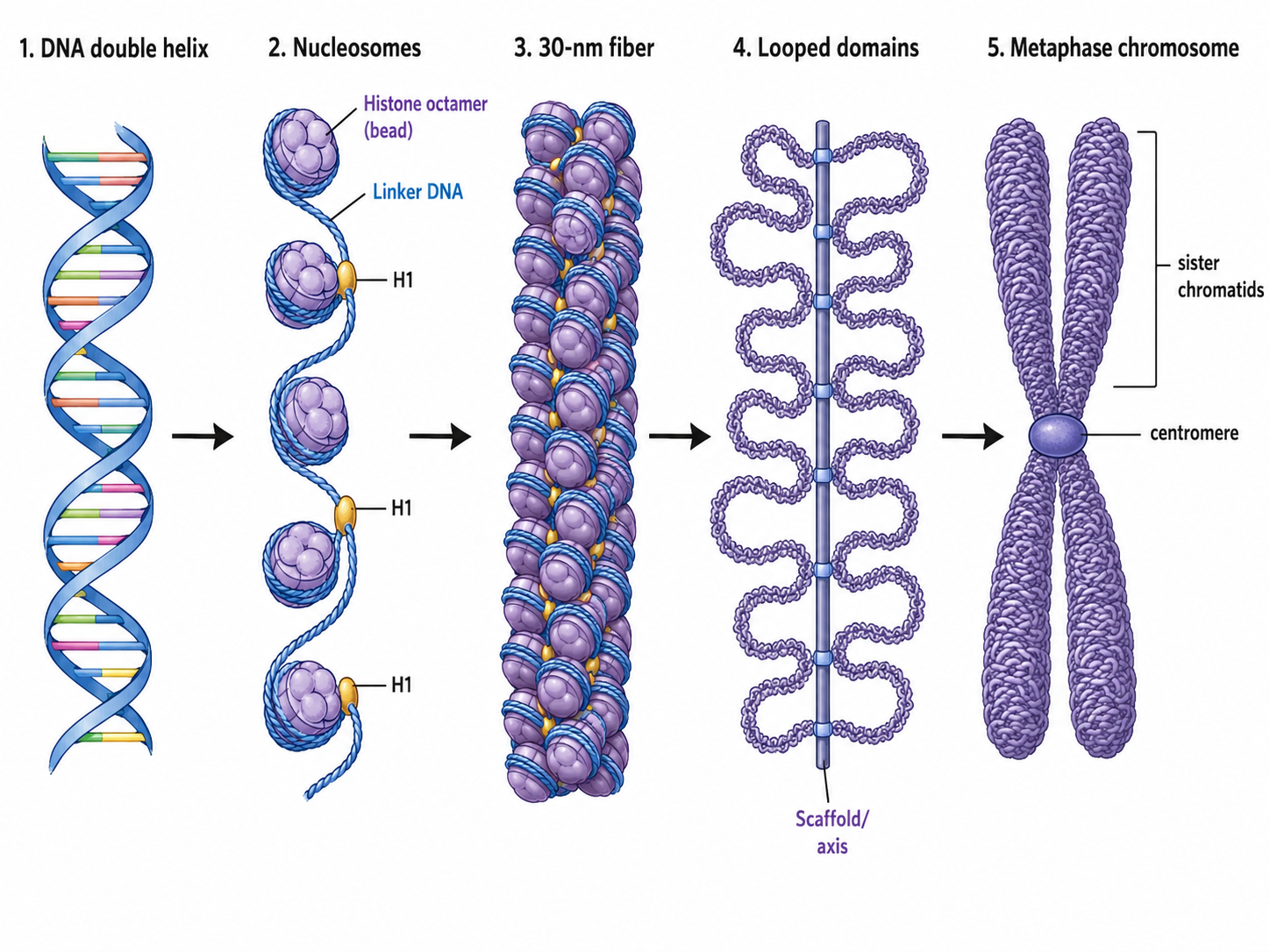
Histone Modifications and Their Functional Consequences
Know the logicHistone tails carry covalent modifications that tune DNA access — the basis of epigenetic control:
- Acetylation (by HATs) neutralizes positive charge on lysines, weakening histone–DNA attraction → loosens chromatin → more transcription. Deacetylation (by HDACs) reverses this → tightens and silences.
- Methylation is context-dependent: it can activate or silence depending on the site.
Quick check: A researcher adds a histone deacetylase (HDAC) inhibitor to cultured cells. Would overall gene expression increase or decrease?
Answer: Increase. Blocking HDACs leaves acetyl groups in place, keeping chromatin open and transcription elevated.
Single Copy vs. Repetitive DNA
Must knowOnly a small fraction of the eukaryotic genome codes for protein; most is noncoding and/or repetitive.
- Single-copy (unique) DNA: present in one or a few copies; this is where most protein-coding genes live, along with regulatory sequences (promoters, enhancers) and introns.
- Repetitive DNA: present in many copies. Tandem repeats are identical units head-to-tail at one site (e.g., satellite DNA at centromeres and telomeres); interspersed repeats are scattered copies largely from transposable elements (mobile DNA).
Quick check: Roughly what fraction of the eukaryotic genome codes for protein, and what makes up most of the rest?
Answer: Only a small fraction codes for protein; most is noncoding — introns, regulatory sequences, and large amounts of repetitive DNA.
Supercoiling
Know the logicB-form DNA has ~10.5 bp per helical turn. Replication or transcription unwinds the strands, creating torsional stress that twists the helix on itself — supercoiling.
- Negative supercoiling (underwound): the predominant cellular state; facilitates strand separation for replication and transcription.
- Positive supercoiling (overwound): accumulates ahead of a moving polymerase and resists unwinding, so it must be relieved.
Topoisomerases manage this: they transiently cut one strand (type I) or both strands (type II), let the strain relax (or introduce supercoils), then reseal the backbone — during packaging, replication, and transcription.
Quick check: During replication, positive supercoils accumulate ahead of the fork. What enzyme relieves this?
Answer: A topoisomerase — it transiently cuts the DNA, lets the strands rotate/pass to relax the strain, then reseals so the fork can advance.
Heterochromatin vs. Euchromatin
Must knowChromatin exists in two broad states, and the distinction is a central gene-regulatory mechanism:
- Euchromatin: loosely packed, accessible, actively transcribed, stains lightly. Most of the working interphase genome.
- Heterochromatin: tightly compacted, transcriptionally inactive, stains darkly. Two types:
- Constitutive: permanently condensed in all cell types; at centromeres and telomeres; gene-poor, rich in satellite repeats.
- Facultative: condensed only in some cells/conditions; classic example = the inactivated X chromosome.
X-Chromosome Inactivation and the Barr Body
Must knowTo equalize X-linked gene dosage, female mammals randomly and permanently inactivate one X per somatic cell early in development, condensing it into facultative heterochromatin visible as the Barr body. Because the choice is random and inherited by all descendants, females are genetic mosaics for X-linked genes.
Quick check: Why is the Barr body facultative rather than constitutive heterochromatin?
Answer: It is conditionally silenced — only one of the two X's, only in certain contexts — not permanently condensed in every cell like centromeric/telomeric (constitutive) heterochromatin.
Telomeres and Centromeres
Telomeres: Protecting Chromosome Ends
Must knowEach linear chromosome ends in a telomere — repetitive DNA (in humans, tandem TTAGGG repeats) plus proteins that cap and protect the ends from degradation, fusion, or being read as a double-strand break.
Telomeres also buffer the end-replication problem: because the lagging strand can't be completed at the very end, chromosomes shorten each division — the telomere erodes instead of essential genes. Telomerase (a reverse transcriptase carrying its own RNA template) extends the repeats and is active in germ cells, stem cells, and most cancers. (Developed further in the DNA Replication guide.)
Centromeres: The Spindle Attachment Point
Must knowThe centromere is the constricted region joining sister chromatids and attaching chromosomes to the spindle. Its roles:
- Kinetochore assembly: the kinetochore (a multiprotein complex) assembles here, and spindle microtubules attach to it to pull chromosomes to the poles in mitosis and meiosis.
- Sister chromatid cohesion: holds chromatids together until anaphase.
- Chromatin: constitutive heterochromatin, rich in tandem satellite repeats.
Quick check: Why is a functional centromere/kinetochore essential for accurate segregation?
Answer: The kinetochore is the attachment point for spindle microtubules. Without it, chromosomes can't be gripped and pulled to the poles, causing missegregation and aneuploidy.
Common Confusions & Tricks
1. The nucleosome octamer — which histones? H1 is not in the octamer; it's the linker histone, outside it. The octamer is 2×(H2A + H2B + H3 + H4). "The core four come in pairs — H1 is the outlier."
2. Acetylation loosens; methylation is context-dependent. Acetylation reliably opens chromatin and promotes transcription; methylation can activate or repress.
3. Euchromatin vs. heterochromatin staining: Euchromatin = light (open, active); heterochromatin = dark (dense, silent). "Hetero = heavy/dark, eu = easy to access."
4. Telomerase is a reverse transcriptase — it makes DNA from its own RNA template; don't equate it with regular DNA polymerase.
5. Positive supercoiling isn't "good." It forms ahead of polymerases and inhibits unwinding; negative supercoiling is the normal state that aids strand separation. Don't read +/− as value judgments.
6. Centromere vs. telomere: both are constitutive heterochromatin, but centromeres are the constricted middle (spindle attachment) and telomeres are the ends (TTAGGG caps).
Key Takeaways
- Nucleosome = histone octamer (2×H2A, 2×H2B, 2×H3, 2×H4) wrapped by DNA. H1 is the linker, not part of the octamer. Histones are basic (positively charged).
- Acetylation loosens chromatin → promotes transcription; deacetylation tightens → silences. Methylation is context-dependent.
- Compaction: "beads on a string" → 30-nm fiber → looped/folded structure → condensed mitotic chromosome.
- Single-copy DNA = most protein-coding genes + regulatory elements; repetitive DNA = tandem repeats + transposable elements; most of the genome is noncoding/repetitive.
- Supercoiling = over-/under-winding; topoisomerases cut and reseal to manage torsional strain. Negative (normal) aids strand separation; positive accumulates ahead of polymerases and is inhibitory.
- Euchromatin = open, active, light-staining; heterochromatin = condensed, silent, dark-staining.
- Constitutive heterochromatin (centromeres, telomeres) is always condensed; facultative is conditional (inactive X = Barr body).
- Telomeres = TTAGGG caps that protect ends and buffer replication-associated shortening.
- Centromeres = constricted region joining sister chromatids and site of kinetochore assembly for spindle attachment.