Priority labels: Must know = cold; Know the logic = mechanism not names; Passage-level = recognize, don't memorize; Optional = skippable.
The Central Dogma: DNA → RNA → Protein
Must knowThe central dogma describes the directional flow of genetic information: DNA is copied into RNA (transcription), and RNA is decoded into protein (translation). DNA is the master archive — the cell makes a working copy (mRNA) and sends it to the ribosome rather than using DNA directly.
Must know the allowed flows and the one that's forbidden:
- DNA → RNA → Protein (the normal path)
- DNA → DNA (replication)
- RNA → DNA (reverse transcription, by retroviruses like HIV using reverse transcriptase)
- Forbidden: protein → nucleic acid. Once information is in amino acid sequence, it cannot be read back.
The most testable application: any DNA mutation can be traced forward — how does the DNA change alter the mRNA codon, and how does that change (or not change) the protein?
Quick check: A cell is treated with a drug that permanently blocks reverse transcriptase. Which type of virus would be most affected, and why?
Answer: Retroviruses (e.g., HIV). They carry an RNA genome and use reverse transcriptase to convert it to DNA that integrates into the host genome. Without the enzyme, the virus cannot establish a permanent infection.
The Triplet Code
Why Triplets?
Know the logicThe code must map a 4-letter alphabet (A, U, G, C) onto 20 amino acids. One nucleotide per amino acid gives only ; two gives — still too few. A triplet code gives , more than enough.
Properties of the Code
Must knowThe four key properties:
- Triplet — each codon is three consecutive mRNA nucleotides specifying one amino acid (or stop).
- Non-overlapping — each nucleotide belongs to exactly one codon (…AUGCGA… reads AUG | CGA).
- Comma-free — no spacer nucleotides; the reading frame is set by the start codon.
- Unambiguous — one codon → one amino acid. (The reverse isn't true: one amino acid can have many codons — degeneracy, below.)
Of the 64 codons, 61 code for amino acids (sense codons) and 3 are stop codons (UAA, UAG, UGA).
Reading a Codon Table
Know the logicThe MCAT supplies a codon table — you read it, never memorize it. Read the mRNA 5'→3', split into non-overlapping triplets starting at the AUG that sets the reading frame, then look up each codon.
Worked example. Given the mRNA:
Find the first AUG (the leading "GC" is 5' UTR, skipped), then read triplets: AUG | GCU | UCA | UAA = Met (start) – Ala – Ser – stop. The peptide is Met–Ala–Ser (the stop codon adds no amino acid). Shifting the start by one nucleotide would change every downstream codon — why setting the frame correctly is essential.
Quick check: If the code were a doublet instead of a triplet, how many amino acids could it encode? Would that be enough?
Answer: , not enough for all 20. A triplet code is the minimum that works.
The Codon–Anticodon Relationship
Must knowTransfer RNA (tRNA) bridges the mRNA codon and the correct amino acid. Two functional sites matter:
- The anticodon, three nucleotides complementary and antiparallel to the mRNA codon.
- The 3' CCA acceptor end, where an aminoacyl-tRNA synthetase covalently attaches the correct amino acid (one synthetase per amino acid — these enzymes do the real "decoding").
The codon (5'→3') pairs antiparallel with the anticodon. If the codon is 5'-AUG-3', the anticodon is 3'-UAC-5' (written 5'-CAU-3'). Positions 1–2 follow standard Watson-Crick rules; position 3 of the codon is the flexible wobble position (next section).
A charged tRNA is an aminoacyl-tRNA. The ribosome catalyzes peptide bond formation; this peptidyl transferase activity lives in the large subunit and is carried out by rRNA — the ribosome is a ribozyme.
Quick check: An mRNA codon reads 5'-GCC-3'. What is the anticodon written in the conventional 5'→3' direction?
Answer: It pairs antiparallel: 5'-GCC-3' pairs with tRNA 3'-CGG-5', written 5'-GGC-3'.
Degenerate Code and Wobble Pairing
Degeneracy
Must knowThe code is degenerate (redundant): most amino acids have more than one codon (synonymous codons). For example, Leu and Ser each have six codons, while Met (AUG) and Trp (UGG) have only one.
Synonymous codons almost always differ at the third (wobble) position.
Wobble Hypothesis
Know the logicAt the third codon position, base-pairing rules relax, so the 5' nucleotide of the anticodon can pair with more than one nucleotide. This is why cells need far fewer than 61 distinct tRNAs. Key examples: inosine (I) in the anticodon pairs with U, C, or A (most flexible); G pairs with both C and U.
Evolutionary implication: degeneracy buffers against mutation. A third-position change often yields a synonymous codon — a silent (synonymous) mutation with no effect on the protein.
Quick check: An mRNA codon 5'-GCU-3' (alanine) mutates to 5'-GCC-3'. What type of mutation is this, and its likely effect?
Answer: GCC also encodes alanine (third position U→C, both synonymous). This is a silent/synonymous mutation; the protein is unchanged.
Missense and Nonsense Codons
Must knowSingle-nucleotide changes have very different consequences depending on what the new codon specifies.
Silent mutation: codon changes, amino acid does not (usually at the wobble position); typically no effect.
Missense mutation: the codon now specifies a different amino acid. Two subtypes by chemical similarity:
- Conservative: similar properties (e.g., Leu → Ile); function usually preserved.
- Non-conservative: chemically different (e.g., charged for nonpolar); more disruptive. Must know the classic example, sickle cell anemia: a single base change in β-globin converts GAG → GUG, swapping glutamate (charged, hydrophilic) for valine (nonpolar, hydrophobic), causing hemoglobin S to polymerize and sickle red cells under low oxygen.
Nonsense mutation: a sense codon becomes a stop codon (UAA, UAG, UGA), creating a premature stop → truncated, usually nonfunctional protein. Generally more severe than missense because all downstream sequence is lost. Stop codons have no tRNAs — they are recognized by release factors.
OptionalTransitions vs. transversions: a transition swaps purine↔purine or pyrimidine↔pyrimidine (ring type preserved); a transversion swaps purine↔pyrimidine (ring type changes). One substitution can be classified both ways — e.g., sickle-cell is a transversion AND a missense mutation.
Frameshift mutations: an insertion or deletion not divisible by 3 shifts the entire reading frame, misreading every downstream codon and usually introducing a premature stop — typically devastating. Indels of multiples of 3 add/remove whole amino acids without frameshifting.
Quick check: A mutation converts UGC (cysteine) to UGA. What type of mutation is this, and the likely effect?
Answer: UGA is a stop codon, so this is a nonsense mutation. Translation terminates prematurely, producing a truncated, almost certainly nonfunctional protein.
Initiation and Termination Codons
The Start Codon: AUG
Must knowTranslation begins at AUG = methionine, which sets the reading frame for everything downstream (the initiator Met is often removed later).
Passage-levelHow the ribosome finds AUG differs by domain:
- Prokaryotes: a Shine-Dalgarno sequence in the 5' UTR base-pairs with the 16S rRNA to position the ribosome; initiator tRNA carries formyl-methionine (fMet).
- Eukaryotes: the ribosome binds the 5' cap and scans to the first AUG (in a favorable Kozak context); initiator tRNA carries regular Met.
Stop Codons: UAA, UAG, UGA
Must knowStop codons have no tRNAs. Protein release factors recognize the stop codon in the A site and trigger release of the completed polypeptide; the ribosome then disassembles (ribosome recycling) and its parts are reused.
OptionalBacterial RF1 reads UAA/UAG, RF2 reads UAA/UGA; eukaryotic eRF1 reads all three.
Quick check: A mutation in the 5' UTR of a prokaryotic mRNA destroys the Shine-Dalgarno sequence. Likely effect on translation?
Answer: The small subunit can't efficiently bind the mRNA, so initiation at the correct AUG is severely reduced or abolished, dropping protein output from that mRNA.
Messenger RNA (mRNA)
Structure of Eukaryotic mRNA
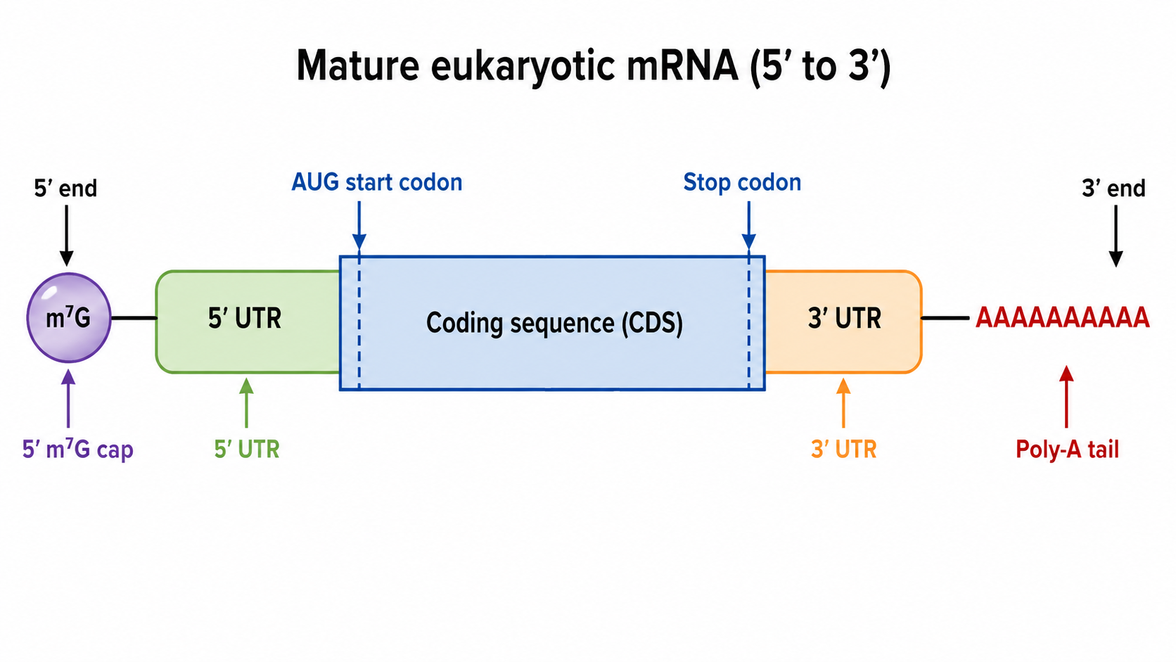
The parts, 5'→3':
- 5' m7G cap: protects from 5' exonucleases, aids nuclear export, and recruits the ribosome via initiation factors.
- 5' UTR: between cap and AUG; not translated; holds regulatory elements (Kozak in eukaryotes).
- Coding sequence (CDS): AUG through the stop codon — the only translated part.
- 3' UTR: downstream of the stop; regulatory (microRNA and RNA-binding-protein sites affecting stability/translation).
- Poly-A tail: ~100–250 adenosines added by poly-A polymerase; protects from 3' exonucleases, aids export, and (via poly-A binding protein) enhances initiation by circularizing the mRNA.
Pre-mRNA Processing (Eukaryotes)
Know the logicThe primary transcript (pre-mRNA) gets three modifications: 5' capping, 3' polyadenylation, and splicing — removal of introns and joining of exons by the spliceosome. Alternative splicing lets one gene produce multiple protein isoforms.
Prokaryotic mRNA: Key Differences
Know the logicProkaryotic mRNAs lack caps and poly-A tails, are often polycistronic (one mRNA, multiple proteins, each with its own SD sequence and AUG) versus monocistronic eukaryotic mRNAs, and — lacking a nucleus — have coupled transcription and translation (ribosomes translate while the mRNA is still being made).
The Universality of the Genetic Code
Know the logicThe code is essentially universal — the same codons mean the same amino acids in nearly all organisms, evidence for common ancestry and the basis for recombinant expression (e.g., human insulin in bacteria). Minor exceptions exist (e.g., mitochondria), but the specifics aren't required.
Quick check: A eukaryotic mRNA has its poly-A tail experimentally removed. Predict two consequences.
Answer: (1) It becomes susceptible to 3' exonucleases, shortening its half-life. (2) Translation efficiency may drop, since poly-A binding protein helps circularize the mRNA and cooperates with the 5' cap to enhance initiation.
Common Confusions & Tricks
1. "Degenerate" ≠ "ambiguous." Degenerate = one amino acid, many codons (many-to-one). Unambiguous = one codon, one amino acid. The code is both at once.
2. Wobble is at the 3' end of the codon but the 5' end of the anticodon. They're antiparallel, so 3' codon ↔ 5' anticodon. Students often reverse the anticodon direction.
3. Stop codons have no tRNA — they have release factors. A common wrong answer is a special "stop tRNA." Release factors (proteins) recognize stops directly.
4. AUG = start AND the only codon for methionine. Every internal Met is AUG, but the ribosome doesn't restart at internal AUGs — context (Kozak/Shine-Dalgarno) and ribosome state determine initiation.
5. The poly-A tail is not encoded in the DNA. It's added post-transcriptionally by poly-A polymerase after cleavage at a polyadenylation signal (AAUAAA) — not a string of T's in the gene.
6. Prokaryotic vs. eukaryotic initiation landmarks: Prokaryote → Shine-Dalgarno + fMet; Eukaryote → 5' cap + scanning + Kozak + regular Met. Don't mix these on bacteria-vs-human questions.
7. Missense vs. nonsense: Missense = wrong amino acid; Nonsense = early stop; Silent = same amino acid. A truncated protein → think nonsense first.
8. The code is "nearly" universal. Strong evidence for common ancestry; a few exceptions (e.g., mitochondria) exist but the reassigned codons aren't required.
9. Frameshifts are their own category — not missense or nonsense — but almost always create a downstream premature stop, sharing the "truncated protein" outcome. ±1 nucleotide causes a frameshift; ±3 does not.
Key Takeaways
Central Dogma
- DNA → RNA → Protein (normal); RNA → DNA allowed (retroviruses); protein → nucleic acid forbidden.
The Triplet Code
- codons; 61 sense + 3 stop (UAA, UAG, UGA)
- Code is triplet, non-overlapping, comma-free, unambiguous, degenerate, and nearly universal
Codon–Anticodon
- mRNA codon (5'→3') pairs antiparallel with tRNA anticodon (3'→5')
- Aminoacyl-tRNA synthetases charge tRNAs; peptidyl transferase is RNA-based (ribosome = ribozyme)
Degeneracy and Wobble
- Most amino acids have multiple synonymous codons, differing at the 3rd (wobble) position
- Inosine (I) in the anticodon pairs with U, C, or A; degeneracy buffers silent mutations
Mutation Types
| Mutation | Codon Change | Amino Acid | Protein Effect |
|---|---|---|---|
| Silent | Changes | Same | Usually none |
| Missense | Changes | Different | Variable (mild to severe) |
| Nonsense | → Stop codon | Truncated | Usually severe |
| Frameshift | Shifts reading frame | All downstream altered | Usually severe |
Initiation and Termination
- Start: AUG (Met/fMet); sets the reading frame
- Prokaryotes: Shine-Dalgarno + fMet-tRNA; Eukaryotes: 5' cap + scanning + Kozak + Met-tRNA
- Stop: UAA, UAG, UGA; recognized by protein release factors (not tRNAs)
mRNA Structure (Eukaryotic)
- 5' m7G cap → 5' UTR → AUG [CDS] Stop → 3' UTR → poly-A tail
- Pre-mRNA processing: capping, polyadenylation, splicing (introns out, exons joined)
- Prokaryotic mRNA: no cap, no poly-A, often polycistronic, transcription-translation coupled
- The genetic code is nearly universal (minor exceptions, e.g., mitochondria — specifics not required)