Nucleic acids store and transmit genetic information. The MCAT tests this topic both conceptually (what does each structural feature do?) and analytically (how does structure predict behavior in denaturation, hybridization, and replication experiments?). Build the model from the smallest unit up.
Priority labels: Must know = cold; Know the logic = mechanism not names; Passage-level = recognize, don't memorize; Optional = skippable.
Nucleotides and Nucleosides
The Building Blocks
Must knowThe fundamental unit is the nucleotide, which has three parts:
- A nitrogenous base (the "letter" of the genetic code)
- A pentose (five-carbon) sugar — deoxyribose in DNA, ribose in RNA
- One or more phosphate groups
A nucleoside is just base + sugar (no phosphate). Add at least one phosphate and it becomes a nucleotide. This matters because many active molecules are nucleotides in free form — ATP is adenosine triphosphate, a nucleotide with three phosphates.
| Term | Components |
|---|---|
| Nucleoside | Base + Sugar (e.g., adenosine) |
| Nucleotide | Base + Sugar + 1–3 phosphates (e.g., AMP, ADP, ATP) |
The base attaches to carbon 1' of the sugar through an N-glycosidic bond (covalent, distinct from the phosphodiester bonds of the backbone).
Quick check: A sample contains "deoxyadenosine." Does this molecule contain a phosphate group?
Answer: No. "Deoxyadenosine" is a nucleoside (deoxyribose + adenine). With phosphate it would be deoxyadenosine monophosphate (dAMP).
Deoxyribose vs. Ribose
Must knowDNA's sugar is 2'-deoxyribose (an H at carbon 2'); RNA's sugar is ribose (an at 2'). Consequences:
- The 2'-OH makes RNA more reactive and less stable — explaining RNA's short lifespan and its catalytic capacity (ribozymes).
- DNA's missing 2'-OH makes the double helix a stable, long-term information archive.
Sugar carbons are numbered with primes (1'–5') to distinguish them from the base atoms. The 3' carbon carries the hydroxyl used in chain elongation; the 5' carbon is where phosphate attaches. These define strand directionality.
Purine and Pyrimidine Bases
Must knowThe bases are aromatic heterocycles in two families:
Purines — fused bicyclic (two rings): Adenine (A), Guanine (G).
Pyrimidines — single six-membered ring: Cytosine (C) (DNA + RNA), Thymine (T) (DNA only; methyl at C5), Uracil (U) (RNA only; like thymine but no C5 methyl).
Mnemonics: "CUT the PY" — Cytosine, Uracil, Thymine are PYrimidines; purines are pure As Gold (A and G).
Know the logicA purine always pairs with a pyrimidine, giving constant helix width. Two purines would bulge; two pyrimidines would narrow.
Quick check: What structural feature distinguishes uracil from thymine?
Answer: Thymine has a methyl group () at the 5-position; uracil has a hydrogen there. Their H-bonding pattern is otherwise identical.
The Sugar-Phosphate Backbone
Phosphodiester Bonds
Must knowNucleotides are linked by phosphodiester bonds: a phosphate bridges the 3'-OH of one sugar to the 5'-carbon of the next.
Know the logic
The bond forms by condensation, releasing pyrophosphate (); hydrolysis of drives the reaction forward. This is why polymerases use triphosphate nucleotides (dNTPs/NTPs) — cleaving two phosphates provides the thermodynamic push.
Strand Polarity
Must knowEvery strand has a 5' end (free phosphate at 5') and a 3' end (free hydroxyl at 3'). By convention, sequences are written 5' → 3'. All polymerases synthesize 5' → 3', adding to the growing 3'-OH end.
The backbone is negatively charged at physiological pH (ionized phosphates). This is why DNA binds positively charged histones in eukaryotes, and why gel electrophoresis separates DNA by size (uniform charge-to-size ratio sends all DNA toward the positive electrode).
Quick check: A dNTP is incorporated by DNA polymerase. What is released, and at which end does incorporation occur?
Answer: Pyrophosphate () is released; incorporation is at the 3' end, extending the chain 5'→3'.
DNA Structure: The Watson-Crick Double Helix
The Model
Must knowThe Watson-Crick model (B-form DNA, the physiological form) describes:
- Two antiparallel strands in a right-handed helix around a common axis
- Sugar-phosphate backbones on the outside, facing water
- Bases stacked inward, shielded from water
- Major and minor grooves from the stacking geometry; the major groove is the primary site for protein-DNA recognition (transcription factors, restriction enzymes)
Antiparallel Orientation
Must know"Antiparallel" means the two strands run in opposite 5'→3' directions. The MCAT will ask you to write a complementary strand accounting for this.
Worked Example: Template → write the complement.
Apply A↔T, G↔C with antiparallel alignment:
Written conventionally (5'→3'):
Base Pairing Specificity and Chargaff's Rules
Must knowChargaff's rules (for any dsDNA):
- and , so purines = pyrimidines in total
- The A+T to G+C ratio varies between species
Base pairing:
- A pairs with T via 2 hydrogen bonds
- G pairs with C via 3 hydrogen bonds
More H-bonds make GC-rich regions more thermally stable.
Passage-levelIndividual H-bonds are weak; the helix's stability comes mainly from cumulative H-bonding plus base-stacking (hydrophobic/van der Waals forces between stacked base pairs). Base stacking is the dominant stabilizing force; H-bonds confer specificity.
Quick check: A dsDNA molecule is 22% guanine. Find A, T, and C.
Answer: [C] = [G] = 22%. [A] = [T] = (100 − 22 − 22)/2 = 28%. So A = 28%, T = 28%, C = 22%.
Function in Transmission of Genetic Information
DNA as the Information Store
Must knowInformation is encoded in the base sequence, not the uniform backbone. The Central Dogma:
The double-stranded structure solves faithful copying: each strand templates a complementary strand during semiconservative replication (Meselson-Stahl). Because pairing is fixed (A–T, G–C), sequence is preserved each round.
Template Function and Strand Terminology
Must knowDuring transcription, one strand is the template strand (= antisense = noncoding). The other, the non-template strand (= coding = sense), matches the mRNA sequence (with U for T). Passages mix these synonyms, so know all three of each.
Quick check: The template strand reads 3'-TACGGGCTA-5'. What is the mRNA?
Answer: mRNA is synthesized 5'→3' complementary to the template, with U for T: 5'-AUGCCCGAU-3'.
DNA Denaturation, Reannealing, and Hybridization
Denaturation
Must knowDenaturation ("melting") separates the two strands into ssDNA. It breaks hydrogen bonds and base stacking, not the covalent phosphodiester backbone. Induced by heat (most tested), extremes of pH, or chemical denaturants (urea, formamide).
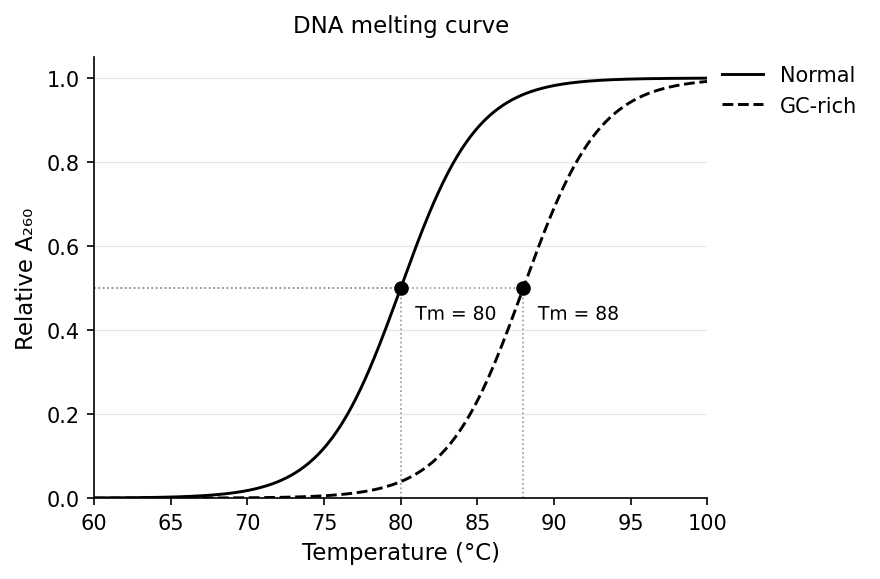
The melting temperature () is where 50% of the DNA is single-stranded. Key trend:
Higher GC → higher (3 H-bonds vs. 2 for AT); longer duplexes also have higher ; AT-rich regions melt first. (Qualitative only — no formula needed.)
The Hyperchromic Effect
Must knowWhen DNA denatures, UV absorbance at 260 nm rises ~30–40% (the hyperchromic effect) because ssDNA absorbs more strongly than dsDNA. Plotting A260 vs. temperature gives a sigmoidal melting curve whose midpoint is ; a GC-rich sample shifts it right.
Reannealing and Hybridization
Must knowSlowly cooling denatured DNA lets complementary strands re-pair into the duplex — reannealing (renaturation). It needs time for complementary strands to collide and slow cooling (rapid "snap cooling" traps ssDNA).
Hybridization is the same process applied to strands from different sources — DNA:DNA, DNA:RNA, or RNA:RNA. It is selective: only sufficiently complementary strands form stable duplexes. This underlies key techniques:
| Technique | Detects |
|---|---|
| Southern blot (probe to DNA) | Specific DNA sequences |
| Northern blot (probe to RNA) | mRNA expression |
| FISH | Chromosomal location / copy number |
| PCR | Amplifies a specific sequence |
| DNA microarray | Genome-wide gene expression |
Stringency (temperature, salt, formamide) controls how many mismatches are tolerated — high stringency requires near-perfect complementarity, low stringency permits mismatches.
Quick check: To detect whether an mRNA is expressed in liver cells, which technique fits and why?
Answer: Northern blot — it separates RNA by size, transfers to a membrane, and uses a complementary probe. (Southern detects DNA.)
Common Confusions & Tricks
1. Nucleoside vs. nucleotide — phosphate is the differentiator. "-side" = no phosphate; "-tide" = phosphate "tied on." Passages name compounds as "adenosine"/"guanosine" — don't assume phosphate is present.
2. Purines vs. pyrimidines. Purines (A, G) are the larger TWO-ring bases; pyrimidines (C, T, U) have ONE ring. Each pair = one big + one small = constant width.
3. GC is stronger, not weaker. G–C = 3 H-bonds, A–T = 2. More bonds = more stable = harder to denature → higher .
4. Antiparallel. Both strands read 5'→3' but in opposite directions. Write the complement 5'→3' by reading the template 3'→5'.
5. Southern vs. Northern. Southern = DNA, Northern = RNA ("D comes before R," DNA is south of RNA).
6. Denaturation breaks H-bonds and stacking, NOT phosphodiester bonds. The covalent backbone stays intact, so reannealing restores the original sequence — which is why hybridization techniques work.
7. Template vs. coding strand. Template = antisense = noncoding (read by RNA polymerase). Coding = sense = non-template (matches mRNA, with T for U). Lock in all three synonyms each.
8. Hyperchromic effect. A260 GOES UP as DNA denatures — higher absorbance = more ssDNA = less double helix.
Key Equations
| Relation | Notes |
|---|---|
| Qualitative only: rises with %GC (3 H-bonds per GC vs. 2 per AT) and with length; AT-rich regions melt first. No formula needed. | |
| Chargaff's rules for dsDNA; used to find base composition from partial data. | |
| If GC% is known, AT% = 100% − GC%, then A% = T% = AT%/2. |