Carbohydrates are the primary fuel molecules of living systems, and the MCAT tests them from two angles: organic chemistry (stereochemistry, ring formation, reactions) and biochemistry (structure-function of biologically important sugars and polymers). Build the mental model of each concept before memorizing names.
Priority labels: Must know = cold; Know the logic = mechanism not names; Passage-level = recognize, don't memorize; Optional = skippable.
Carbohydrate Classification: The Big Picture
Must knowA carbohydrate (saccharide, sugar) is a polyhydroxy aldehyde or polyhydroxy ketone, or a compound hydrolyzable into one. The name comes from the empirical formula ("hydrates of carbon").
Aldoses vs. Ketoses
Must knowThe carbonyl position defines the two families:
- Aldoses carry the carbonyl as an aldehyde at C1 (glucose, galactose).
- Ketoses carry it as a ketone, usually at C2 (fructose).
Glucose is an aldohexose; fructose is a ketohexose.
Classification by Carbon Chain Length
Must knowMonosaccharides are also named by carbon count: triose (3), tetrose (4), pentose (5), hexose (6). High-yield members: glyceraldehyde and DHAP (trioses central to glycolysis), ribose/deoxyribose (pentoses), glucose/galactose/fructose (hexoses).
Quick check: Is fructose an aldose or ketose, and how many carbons does it have?
Answer: Fructose is a ketose with 6 carbons — a ketohexose. Its carbonyl is at C2, whereas glucose's is at C1.
Nomenclature, Common Names, and Absolute Configuration
Stereochemistry Recap (full treatment in the 5D Carbohydrates guide)
Must knowStereochemistry is read from Fischer projections (chain vertical, most oxidized carbon on top). D/L is set by the highest-numbered chiral center (C5 in glucose): on the right = D, on the left = L. Nearly all biological sugars are D, referenced to D-glyceraldehyde. D/L is a family label, distinct from R/S (CIP, per-center) and from optical rotation — D does not imply R or dextrorotatory. For Fischer-to-R/S assignment and absolute configurations, see the 5D guide.
Common Names Worth Knowing
Must knowGlucose = dextrose (+); fructose = levulose (−); galactose = C4 epimer of glucose; mannose = C2 epimer of glucose; ribose = RNA pentose; deoxyribose = DNA pentose, lacks the C2-OH.
Quick check: D-fructose is called levulose. Does this mean fructose is an L-sugar?
Answer: No. "Levo-" refers to optical rotation (rotates light left), not D/L configuration. D-fructose is still a D-sugar by its C5 center; optical rotation and D/L are independent.
Cyclic Structure and Conformations of Hexoses
Cyclization Recap (full treatment in the 5D Carbohydrates guide)
Must knowIn solution the carbonyl reacts intramolecularly with a hydroxyl, forming a cyclic hemiacetal (aldose) or hemiketal (ketose); ring closure creates the new chiral center called the anomeric carbon. Glucose's C1 aldehyde + C5 OH gives a six-membered pyranose; fructose's C2 ketone + C5 OH gives a five-membered furanose (the form in sucrose). A Haworth projection is the planar side view (Fischer "right" → "below," up for D-sugars); the real geometry is a chair. β-D-glucose is the most stable monosaccharide because in its chair all five non-H substituents are equatorial — biochemically relevant because it makes glucose the dominant cellular fuel and makes β-1,4-linked cellulose rigid. See the 5D guide for Haworth-to-chair conversion details.
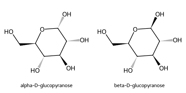
Quick check (worked example): In a Haworth of β-D-glucose, is the anomeric OH above or below? Convert to a chair: axial or equatorial?
Answer: Above the ring (cis to C6). In the chair this is equatorial — exactly why β-D-glucose is more stable than the α anomer.
Epimers, Anomers, and Mutarotation
Must knowRecap (full treatment in the 5D Carbohydrates guide): Epimers differ at exactly one chiral center — glucose/galactose are C4 epimers, glucose/mannose are C2 epimers. Anomers are the special case differing only at the anomeric carbon (C1 in aldoses, C2 in fructose): α = anomeric OH axial (below ring in D-glucose Haworth), β = equatorial (above). Mutarotation is the interconversion of α and β through the open-chain form; dissolving a pure anomer in water shifts its optical rotation to the equilibrium value (~36% α, 64% β). The MCAT tests the direction of the change, not the numbers.
Quick check: You dissolve pure α-D-glucose, the more strongly dextrorotatory anomer, in water. Does the rotation increase or decrease, and why?
Answer: It decreases, because α converts to the less dextrorotatory β via the open-chain form until the equilibrium mixture is reached.
Glycosidic Bonds and Reducing Sugars
Glycosidic Linkages Recap (full mechanism in the 5D Carbohydrates guide)
Must knowWhen the anomeric carbon condenses with an of another molecule, the hemiacetal becomes an acetal (a glycoside), releasing water. A linkage is named by anomeric configuration (α/β) and the carbon numbers joined — e.g., α(1→4) is anomeric C1 of the donor bonded to C4 of the acceptor. Once the glycoside forms the anomeric configuration is locked and mutarotation stops. Metabolically, these bonds are stable at physiological pH and are cleaved by stereospecific glycosidases: lactase (β-galactosidase) cleaves lactose's β(1→4), α-amylase cleaves starch's α(1→4). That stereospecificity is why lactase deficiency causes lactose intolerance — the body simply has no enzyme for the β bond.
Reducing vs. Non-Reducing Sugars
Must knowA reducing sugar has a free anomeric carbon that can open to an aldehyde (or α-hydroxy ketone) and reduce an oxidizing agent. All monosaccharides are reducing. A disaccharide is reducing only if one anomeric carbon is free.
- Reducing: maltose, lactose, cellobiose.
- Non-reducing: sucrose (both anomeric carbons locked).
Benedict's and Fehling's tests use (blue) reduced to (brick-red); Tollens' deposits a silver mirror (). All detect a free anomeric carbon, so they agree. Sucrose tests negative.
Worked example: Benedict's tests on glucose, sucrose, a 50:50 glucose/sucrose mix, and fructose — which are positive?
- Glucose: positive (free anomeric carbon).
- Sucrose: negative (both anomeric carbons locked).
- 50:50 mix: positive (glucose reacts; sucrose doesn't interfere).
- Fructose: positive — under basic conditions the α-hydroxy ketone tautomerizes to an enediol, reducing .
Oxidation/Reduction Products Recap (full treatment in the 5D Carbohydrates guide)
Passage-levelOxidizing the C1 aldehyde gives an aldonic acid (glucose → gluconic acid; the Benedict's/Tollens' reaction); oxidizing the C6 gives a uronic acid (glucose → glucuronic acid, the conjugate used in hepatic glucuronidation to clear drugs and bilirubin); oxidizing both ends gives an aldaric acid; reducing the carbonyl gives an alditol (glucose → sorbitol).
Monosaccharides: Key Members and Their Roles
Must knowKnowing each sugar's structural relationship to glucose prevents confusion.
- Glucose — central fuel; phosphorylated to glucose-6-phosphate to trap it in the cell, then enters glycolysis, the pentose phosphate pathway, or glycogen.
- Galactose — C4 epimer of glucose, from lactose; converted to glucose-1-phosphate via the galactose metabolism pathway. Passage-level GALT deficiency causes classic galactosemia.
- Fructose — ketohexose (C2 carbonyl); metabolized in the liver via fructose-1-phosphate. Optional aldolase B deficiency → hereditary fructose intolerance.
- Mannose — C2 epimer of glucose; key in N-linked glycosylation (mannose-6-phosphate targets enzymes to lysosomes).
- Ribose / deoxyribose — ribose is the pentose in RNA, ATP, NAD(H), FAD, CoA; deoxyribose lacks the C2-OH and is the DNA backbone.
Quick check: Why is RNA hydrolyzed by base but DNA is not?
Answer: RNA's C2-OH acts as an intramolecular nucleophile, attacking the adjacent phosphate (via a 2',3'-cyclic phosphate) to cleave the backbone. DNA lacks the C2-OH, so this mechanism is unavailable, making it far more base-stable.
Disaccharides
Must knowTwo monosaccharides joined by one glycosidic bond. Know these cold:
| Disaccharide | Linkage | Components | Reducing? | Notes |
|---|---|---|---|---|
| Maltose | α(1→4) | Glc + Glc | Yes | from starch; cleaved by maltase |
| Lactose | β(1→4) | Gal + Glc | Yes | milk sugar; cleaved by lactase |
| Sucrose | α(1→β2) | Glc + Fru | No | both anomeric carbons locked |
| Cellobiose | β(1→4) | Glc + Glc | Yes | repeat unit of cellulose; indigestible |
Quick check: Why does sucrose test negative on Benedict's while maltose tests positive, even though both are disaccharides?
Answer: In maltose only one anomeric carbon is in the bond; the other (C1 of the second glucose) is free and can open to an aldehyde. In sucrose both anomeric carbons (C1 of glucose, C2 of fructose) are locked — no free anomeric carbon, no reducing activity.
Polysaccharides
Must knowLong polymers of monosaccharides. The key idea: bond type determines function.
Starch (Amylose + Amylopectin)
Must knowPlant storage polysaccharide:
- Amylose: linear glucose, α(1→4) linkages; coils into a helix.
- Amylopectin: α(1→4) backbone with α(1→6) branch points (~every 24–30 residues).
Salivary/pancreatic α-amylase cleaves internal α(1→4) bonds; isomaltase cleaves α(1→6) branches.
Glycogen
Must knowAnimal storage polysaccharide (liver, muscle): same α(1→4) backbone + α(1→6) branches as amylopectin but more branched (~every 8–12 residues). Know the logic more branches → more non-reducing ends → faster glucose mobilization by glycogen phosphorylase, which acts at non-reducing ends and releases glucose-1-phosphate by phosphorolysis (using , not water).
Cellulose
Must knowPlant structural polysaccharide: linear glucose with β(1→4) linkages. Adjacent units flip 180°, enabling hydrogen bonding between chains → rigid, insoluble fibers. Humans lack cellulase, so cellulose is dietary fiber.
Chitin
Passage-levelLinear polymer of N-acetyl-D-glucosamine with β(1→4) bonds; structural in fungal walls and arthropod exoskeletons; indigestible to humans.
The key insight: α vs. β linkage decides whether a glucose polymer is digestible (starch/glycogen, α) or indigestible (cellulose, β), despite the same monomer.
Quick check: Amylose and cellulose are both linear glucose polymers. Why does amylose form a helix while cellulose forms flat sheets?
Answer: Amylose's α(1→4) linkages bend the chain into a helix. Cellulose's β(1→4) linkages flip each glucose 180°, giving a flat, extended chain that hydrogen-bonds laterally into rigid sheets.
Common Confusions & Tricks
1. D/L vs. R/S vs. optical rotation — three independent systems. D does not mean R at all centers, and does not mean dextrorotatory. Fructose is D (by C5) yet levorotatory (−). Keep them in separate boxes.
2. α means "axial" for D-sugars in the chair — but only at the anomeric carbon. It does NOT mean all axial substituents are α.
3. Epimers vs. anomers: the anomeric carbon is the divider. Both differ at one center; an anomer differs specifically at the anomeric carbon. Galactose is the C4 epimer of glucose, not an anomer.
4. Sucrose is the only common non-reducing disaccharide (both anomeric carbons locked). A non-sucrose disaccharide is almost certainly reducing.
5. Branch density: "Glycogen → Greater branching → Glucose released faster." Amylopectin branches less often (~24–30).
6. β(1→4) = "can't digest without special enzymes." Cellulose and chitin both use β(1→4); humans digest neither.
7. Mutarotation requires a free anomeric OH — glycosides cannot mutarotate.
8. The "right = below" Haworth rule fails at the ring-closure carbon. For C5 of D-glucopyranose, (C6) goes above even though it's on the right in Fischer, because C5's oxygen is incorporated into the ring. Don't apply right→below to the carbon that closes the ring.
9. Positive Fehling's/Benedict's → reducing sugar; negative → sucrose (or a full acetal/glycoside).
10. Lactose hydrolysis by acid vs. lactase: acid can hydrolyze the β(1→4) bond, but lactase is stereospecific for β-linkages; the intestine isn't acidic enough for fast non-enzymatic hydrolysis, so lactase deficiency means undigested milk sugar.
Key Equations
| Equation / Relationship | When to Use |
|---|---|
| Empirical formula of carbohydrates; = number of carbons. | |
| = chiral centers. An aldohexose has 4 → (8 D + 8 L). | |
| Mutarotation: ~36% α, ~64% β, <1% open-chain. | |
| Benedict's/Fehling's: reducing sugar reduces (blue) to (brick-red). Negative for sucrose. | |
| Glycoside formation (dehydration); reverse = acid hydrolysis. | |
| Phosphorolysis of α(1→4) bonds (not hydrolysis); product is glucose-1-phosphate. |