Priority labels: Must know = cold; Know the logic = mechanism not names; Passage-level = recognize, don't memorize; Optional = skippable.
What Makes a Cell "Eukaryotic"?
Must knowThe core idea is compartmentalization: a eukaryotic cell divides its interior into membrane-enclosed rooms, each with its own chemical environment, enzymes, and job. This is the conceptual thread running through the whole guide.
The three defining hallmarks of eukaryotes:
- A membrane-bound nucleus housing the genome
- Membrane-bound organelles that compartmentalize functions
- Division by mitosis (meiosis for gametes), vs. binary fission in prokaryotes
Eukaryotes are also larger than prokaryotes and have linear chromosomes wrapped on histones plus a cytoskeleton.
Quick check: A bacterium produces ATP at its plasma membrane and has ribosomes, but no nucleus. Why is it not eukaryotic?
Answer: It lacks a membrane-bound nucleus and membrane-bound organelles; its DNA floats in the cytoplasm (nucleoid) and its ribosomes are 70S, not 80S.
The Nucleus: Command Center of the Cell
Why Compartmentalize the Genome?
Know the logicSequestering DNA in the nucleus physically separates transcription (DNA → RNA, in the nucleus) from translation (RNA → protein, in the cytoplasm). This lets mRNA be processed (capping, polyadenylation, splicing) before reaching a ribosome. Prokaryotes, lacking a nucleus, translate mRNA while it is still being transcribed.
The Nuclear Envelope and Nuclear Pores
Must knowThe nuclear envelope is a double membrane continuous with the rough ER — so the perinuclear space is continuous with the rER lumen, and the outer membrane bears ribosomes.
Nuclear pores penetrate the envelope and act as selective gatekeepers: small molecules diffuse through, but large proteins and RNA require signal-mediated transport. Nuclear proteins carry a nuclear localization signal (NLS) recognized by import receptors; processed RNAs are exported outward.
Chromatin and Genetic Storage
Must knowDNA is organized as chromatin (DNA wrapped on histones into nucleosomes). It exists as loosely packed euchromatin (active) or tightly packed heterochromatin (silent), and condenses into visible chromosomes during division. Human somatic cells have 46 chromosomes; the genome is stored in the nucleus (small exception: mtDNA).
The Nucleolus
Must knowThe nucleolus is a dense, non-membrane-bound region inside the nucleus dedicated to ribosome biogenesis: rRNA transcription, rRNA processing, and assembly of the ribosomal subunits (60S + 40S) that are then exported to the cytoplasm. Cells making many proteins have prominent nucleoli.
Quick check: A student claims the nucleolus is where mRNA is synthesized. What is wrong with this?
Answer: mRNA is made throughout the nucleus by RNA polymerase II. The nucleolus is dedicated to rRNA (RNA pol I) and ribosomal subunit assembly — not mRNA.
Mitochondria: The Powerhouse of the Cell
Structure: Two Membranes, Two Compartments
Must knowThe double membrane creates four regions:
- Outer membrane (OMM): smooth; porins make it permeable to small molecules and ions.
- Intermembrane space (IMS): ionic composition resembles cytoplasm, except protons () are pumped here by the ETC, creating the proton gradient.
- Inner membrane (IMM): impermeable, folded into cristae (increase surface area); holds the electron transport chain (Complexes I–IV) and ATP synthase.
- Matrix: innermost; holds the citric acid (Krebs) cycle, β-oxidation enzymes, mitochondrial 70S ribosomes, and mitochondrial DNA (mtDNA).
Site of ATP Production
Must knowThe mitochondrion is the primary site of ATP production in aerobic cells.
| Pathway | Location |
|---|---|
| Citric acid cycle, β-oxidation, pyruvate → acetyl-CoA | Matrix |
| Electron transport chain + ATP synthase | Inner membrane |
Note: glycolysis occurs in the cytoplasm, not the mitochondrion.
Self-Replication and Endosymbiotic Theory
Must knowMitochondria have their own circular DNA, 70S ribosomes (bacterial-type), and reproduce by binary fission. With their double membrane and maternal inheritance, this is the evidence for the endosymbiotic theory: mitochondria descend from a free-living bacterium engulfed by an ancestral host cell. (Chloroplasts in plants show the same pattern.)
Quick check: A drug inhibits 70S ribosomes. Would it affect protein synthesis in the mitochondrial matrix?
Answer: Yes. Mitochondria contain 70S ribosomes (bacterial origin) that make a subset of mitochondrial proteins — so the drug impairs that synthesis. (This is why some antibiotics, e.g. chloramphenicol, have mitochondrial side effects.)
Ribosomes: The Protein Synthesis Machine
Must knowEukaryotic ribosomes are 80S (60S + 40S subunits) and occur in two places:
- Free ribosomes (cytoplasm) — make cytoplasmic, nuclear, mitochondrial, and peroxisomal proteins.
- Membrane-bound ribosomes (on rough ER) — make secreted, membrane, and lysosomal proteins.
The signal sequence on the growing polypeptide decides the fate (see ER section).
Quick check: A ribosome is translating a cytoplasmic enzyme that will never leave the cell. Free or membrane-bound?
Answer: Free. Ribosomes only associate with the rER when the nascent polypeptide carries a signal sequence; cytoplasmic proteins lack it.
Endoplasmic Reticulum: The Cell's Biosynthetic Highway
Must knowThe endoplasmic reticulum (ER) is a network of membrane sacs and tubules continuous with the outer nuclear membrane; its lumen is a distinct compartment. Two functional regions:
Rough Endoplasmic Reticulum (rER)
Must knowThe rough ER is studded with ribosomes and is the entry point of the secretory pathway. Proteins destined for secretion, membranes, or lysosomes carry an N-terminal signal sequence that targets the ribosome to the rER, threading the polypeptide into the lumen; proteins without it stay in the cytosol.
Functions: synthesis of secreted, transmembrane, and lysosomal proteins; N-linked glycosylation; initial folding/quality control; and synthesis of membrane phospholipids.
Smooth Endoplasmic Reticulum (sER)
Must knowThe smooth ER lacks ribosomes. Its functions vary by cell type:
- Lipid and steroid hormone synthesis — prominent in steroidogenic cells (adrenal cortex, gonads)
- Detoxification — liver hepatocytes (cytochrome P450 enzymes)
- Calcium storage — in muscle, the specialized sarcoplasmic reticulum stores/releases for contraction
Reference: hepatocyte glycogen metabolism (glucose-6-phosphatase) also occurs in the sER.
Quick check: A mutation removes the N-terminal signal sequence from a normally secreted protein. Where does it end up, and why?
Answer: Made on a free ribosome and stuck in the cytosol — without the signal sequence the ribosome never reaches the rER, so the protein never enters the secretory pathway.
Golgi Apparatus: The Cell's Postal System
Structure
Must knowThe Golgi apparatus is a polarized stack of flattened cisternae with two faces:
- Cis face: "receiving" side facing the ER; gets vesicles from the rER.
- Trans face: "shipping" side facing the plasma membrane; dispatches processed proteins in vesicles.
Proteins move cis → medial → trans.
Functions
Must knowThe Golgi is the cell's sorting and modification station:
- Further glycosylation of proteins (trimming/extending sugars)
- Proteolytic processing of some proproteins/prohormones into active forms
- Sorting and packaging at the trans face into vesicles bound for the plasma membrane, lysosomes, or storage granules
The Secretory Pathway: Putting It All Together
Must knowThe order:
Free ribosome → signal sequence → rER → transport vesicles → Golgi (cis → trans) → secretory vesicles → exocytosis at plasma membrane
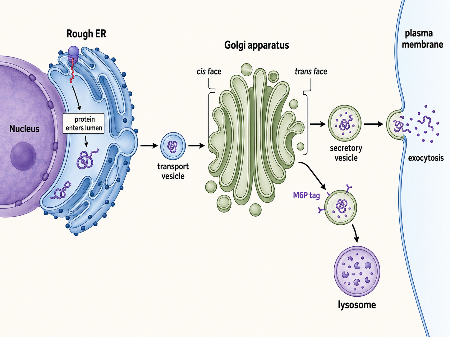
For lysosomal proteins, the pathway diverges at the trans-Golgi: a mannose-6-phosphate (M6P) tag routes the enzymes to lysosomes instead of the plasma membrane.
Quick check: A drug blocks all vesicle fusion with the Golgi cis face. Which proteins most directly accumulate near the Golgi?
Answer: Newly synthesized proteins from the rER, in transport vesicles, accumulate — they can't be received by the cis-Golgi. The secretory pathway halts at the rER-to-Golgi step.
Lysosomes: The Cell's Recycling Center
Structure and Contents
Must knowLysosomes are membrane-bound vesicles of acid hydrolases (proteases, lipases, nucleases, glycosidases) that work best at an acidic pH (~4.5–5.0), maintained by a proton pump (V-type ATPase) that pumps into the lumen.
Functions
Must knowLysosomes degrade material from three sources:
- Autophagy — digesting the cell's own damaged organelles (enclosed in an autophagosome that fuses with a lysosome)
- Phagocytosis — engulfed pathogens/debris (phagosome fuses with lysosome)
- Endocytosis — internalized extracellular material (e.g., LDL) routed via endosomes to lysosomes
Because they are packed with hydrolases, lysosomes are called "suicide bags": enzyme release contributes to apoptosis.
Lysosomal Storage Diseases
Passage-levelDeficiency of a specific lysosomal hydrolase causes substrate to accumulate, producing lysosomal storage diseases (e.g., Tay-Sachs, hexosaminidase A deficiency; Gaucher, glucocerebrosidase deficiency). The lesson: one organelle's failure cascades to the whole cell.
Quick check: Why does lysosomal dysfunction in Tay-Sachs hit neurons so hard, even though all cells have lysosomes?
Answer: Neurons have high ganglioside turnover and are post-mitotic — they can't divide to dilute or replace the accumulating material, so it builds progressively, causing neurodegeneration.
Peroxisomes: Managing Reactive Oxygen Species
Structure and Function
Must knowPeroxisomes are small, single-membrane organelles (abundant in liver/kidney) that both generate and destroy hydrogen peroxide (). The signature enzyme catalase converts toxic into water and oxygen:
Major Functions
Must know- β-oxidation of very-long-chain fatty acids (VLCFAs) — peroxisomes shorten chains too long for mitochondria (regular β-oxidation is mitochondrial)
- Plasmalogen synthesis (a phospholipid important in myelin)
- Detoxification (e.g., liver)
Biogenesis
Must knowUnlike mitochondria, peroxisomes have no DNA. Their proteins are nuclear-encoded and imported post-translationally via a peroxisomal targeting signal (PTS), bypassing the ER/Golgi.
Passage-level Zellweger syndrome is a peroxisomal biogenesis disorder causing VLCFA accumulation.
Quick check: Newly synthesized peroxisomal proteins bypass the ER and Golgi and go directly to peroxisomes. What feature allows this?
Answer: A peroxisomal targeting signal (PTS) recognized by cytoplasmic receptors that escort the protein directly to the peroxisome, bypassing the secretory pathway.
Other Canonical Eukaryotic Cell Structures
Cytoskeleton
Must knowThe cytoskeleton gives cells shape, enables movement, and organizes organelles. Three components:
| Filament | Monomer | Functions |
|---|---|---|
| Microfilaments (actin) | G-actin | Shape, muscle contraction, cytokinesis, motility |
| Intermediate filaments | various (keratin, lamins) | Mechanical strength, nuclear lamina |
| Microtubules | α/β-tubulin | Transport (kinesin/dynein), mitotic spindle, cilia/flagella |
Motor proteins: kinesin moves cargo toward the (+) end (toward the periphery); dynein moves toward the (−) end (toward the nucleus).
Centrosome and Centrioles
Must knowThe centrosome is the main microtubule-organizing center (MTOC) of animal cells; it contains two centrioles (9 triplets, 9+0) and organizes the mitotic spindle. Centrioles also form basal bodies for cilia and flagella, which have a 9+2 axoneme (9 outer doublets + 2 central singlets) powered by dynein.
Plasma Membrane and Cell Coat
Must knowThe plasma membrane is a phospholipid bilayer with embedded proteins and cholesterol, which buffers membrane fluidity across temperatures. Its extracellular face carries the carbohydrate glycocalyx for cell recognition, adhesion, and signaling.
Vacuoles
Passage-levelVacuoles store material. The large plant central vacuole maintains turgor pressure; animal cells have smaller vacuoles.
Quick check: Taxol stabilizes microtubules, preventing depolymerization. Why does this kill rapidly dividing cancer cells?
Answer: Mitosis needs dynamic microtubule polymerization/depolymerization to build and disassemble the spindle. Stabilized microtubules can't depolymerize, so chromosomes can't segregate, division arrests, and apoptosis follows — hitting rapidly dividing cells hardest.
Common Confusions & Tricks
1. Mitochondria have 70S ribosomes — not the 80S cytoplasmic ones. They are bacterial in origin (endosymbiotic theory). Antibiotics targeting 70S ribosomes can affect mitochondria.
2. The nucleolus makes rRNA and assembles ribosomal subunits — NOT mRNA. "Involved in protein synthesis" is a trap; it's specifically ribosome biogenesis.
3. Rough ER vs. Smooth ER by function. rER: ribosomes → protein secretion + membrane biogenesis. sER: no ribosomes → lipid/steroid synthesis, detox (liver), Ca²⁺ storage (muscle). Steroid-secreting cells → abundant sER.
4. Peroxisomes ≠ Lysosomes. Lysosomes: acid hydrolases, degrade debris, derive from the Golgi (M6P). Peroxisomes: oxidases + catalase, handle fatty acids and , do NOT come from the secretory pathway.
5. The secretory pathway order — know it cold. Ribosome → rER → vesicles → Golgi (cis → trans) → secretory vesicles → plasma membrane (or lysosomes via M6P). Expect "defect at step X → where do proteins accumulate?"
6. ER continuity = nuclear envelope. The outer nuclear membrane is continuous with the rER, so the perinuclear space is continuous with the rER lumen.
7. Mitochondrial DNA is maternally inherited. Sperm mitochondria are destroyed after fertilization.
8. Peroxisomes have NO DNA — unlike mitochondria. Don't overextend the endosymbiotic theory.
9. β-oxidation location: mitochondria for regular fatty acids, peroxisomes for VLCFAs. VLCFA accumulation → peroxisomal dysfunction.
10. Lysosomal pH is ~4.5–5, cytoplasm ~7.2. A V-ATPase acidifies the lumen; acid hydrolases are relatively inactive if released into the neutral cytoplasm (a safety feature).
Key Takeaways
Defining Eukaryotic Features
- Membrane-bound nucleus, membrane-bound organelles, 80S cytoplasmic ribosomes, divide by mitosis
- Key advantage: compartmentalization allows specialized biochemical environments
Nucleus
- Double-membrane nuclear envelope continuous with rER; nuclear pores for selective transport (NLS)
- Nucleolus: non-membrane-bound; rRNA transcription + ribosomal subunit (60S + 40S) assembly
Mitochondria
- Double membrane: outer (permeable, porins), inner (impermeable, cristae, ETC + ATP synthase), IMS, matrix
- Matrix: Krebs cycle, β-oxidation, mtDNA, 70S ribosomes; protons pumped to IMS flow back through ATP synthase
- Self-replicating, maternally inherited mtDNA → endosymbiotic origin
ER
- rER: ribosomes; secreted/membrane protein synthesis; N-linked glycosylation; phospholipid synthesis
- sER: no ribosomes; steroid/lipid synthesis; detox (liver); Ca²⁺ storage (sarcoplasmic reticulum)
Golgi Apparatus
- Cis (receives from ER) → trans (dispatches to plasma membrane, lysosomes, storage)
- Glycosylation, proteolytic processing, sorting; M6P tag routes enzymes to lysosomes
Lysosomes
- Single membrane; acid hydrolases; pH ~4.5–5 via V-ATPase
- Degrade material from phagocytosis, endocytosis, autophagy
- Storage diseases (Tay-Sachs, Gaucher) = hydrolase deficiency → substrate accumulation
Peroxisomes
- Single membrane; oxidases + catalase; manage
- VLCFA β-oxidation, plasmalogen synthesis, detox
- No DNA; proteins imported via PTS signals; defects → Zellweger syndrome
Secretory Pathway Summary
Cytoskeleton
- Actin microfilaments: shape, motility, cytokinesis
- Intermediate filaments: mechanical stability, nuclear lamina
- Microtubules: transport (kinesin = +end; dynein = −end), mitotic spindle, cilia (9+2), centrioles (9+0)