The cytoskeleton is one of those topics where understanding the logic makes everything click. Think of the cell as a city: it needs structural scaffolding to hold its shape, highways for shipping cargo, and contractile machinery for movement and division. There are three distinct systems — microfilaments, microtubules, and intermediate filaments — each with its own composition, dynamics, and roles. Master those distinctions and you have this topic.
Priority labels: Must know = cold; Know the logic = mechanism not names; Passage-level = recognize, don't memorize; Optional = skippable.
General Function in Cell Support and Movement
Must knowThe cytoskeleton is a dynamic, three-dimensional network of protein filaments in the cytoplasm of eukaryotic cells. It serves three broad functions:
- Mechanical support — resists deformation, maintains cell shape, anchors organelles.
- Intracellular transport — tracks along which motor proteins carry cargo.
- Cell and organelle movement — muscle contraction, amoeboid crawling, chromosome segregation, cytokinesis.
Critically, it is not static: filaments continuously polymerize and depolymerize, letting the cell rapidly reorganize. This dynamic quality — not just the presence of filaments — is what enables movement and division.
(Prokaryotes have homologs — MreB ~ actin, FtsZ ~ tubulin — but the MCAT focuses on eukaryotes.)
Quick check: A cell treated with a drug that permanently freezes all cytoskeletal filaments in their polymerized state would be unable to do what?
Answer: Cytokinesis, chromosome segregation (the spindle must elongate and shorten), and directed migration — all require filament dynamics, not just the presence of filaments.
Microfilaments: Composition and Role in Cleavage and Contractility
Composition and Structure
Must knowMicrofilaments (actin filaments) are the thinnest system (~7 nm). They are helical polymers of globular G-actin; polymerized, they form the two-stranded F-actin helix. Each G-actin binds ATP, hydrolyzed to ADP after incorporation — the energy governs filament dynamics, not direct mechanical work.
Actin filaments are polar: the barbed (plus, +) end grows faster; the pointed (minus, −) end loses monomers. Treadmilling is the key consequence — at steady state, monomers add at the barbed end at the same rate they leave the pointed end, so the filament "moves" without changing length. This drives cell crawling: the leading edge extends lamellipodia and filopodia by actin polymerization at the membrane.
Role in Contractility and Cytokinesis
Must knowActin plus the motor myosin II generates contractile force. In dividing animal cells they form the contractile ring — a transient belt of actin + myosin II beneath the membrane at the cell equator. Myosin walks along actin, constricting the ring like a purse-string and pinching the cell into two via the cleavage furrow. This is why microfilaments are essential for cytokinesis: block actin and you get a binucleate cell that fails to divide. Actin–myosin also underlies muscle contraction, migration, phagocytosis, and endocytosis.
Passage-levelCytochalasin caps barbed ends and inhibits actin polymerization; phalloidin stabilizes F-actin (research tool).
Quick check
A researcher treats dividing cells with cytochalasin. Mitosis proceeds normally, but the cells end up with two nuclei in a single cell body. Why?
Answer: Cytochalasin blocks actin polymerization, so the contractile ring cannot form. Mitosis (microtubule-dependent) completes, but cytokinesis fails with no ring to make a cleavage furrow.
Microtubules: Composition and Role in Support and Transport
Composition and Structure
Must knowMicrotubules are the largest filaments (~25 nm outer diameter): hollow tubes of α-/β-tubulin heterodimers stacked head-to-tail into protofilaments (13 per tube). Each dimer binds GTP; hydrolysis to GDP drives dynamics. They are polar — the fast-growing plus end points away from the MTOC, the minus end is anchored at the MTOC.
Know the logicDynamic instability: individual microtubules switch between rapid growth and rapid shrinkage (catastrophe), with occasional rescue. A "GTP cap" at the plus end stabilizes the filament; lose the cap and it rapidly depolymerizes. This lets the cell quickly remodel its microtubule network.
Role in Intracellular Transport
Must knowTwo motor families move cargo along microtubules:
| Motor | Direction | Key Roles |
|---|---|---|
| Kinesin | toward + end (anterograde, outward) | vesicle transport to periphery |
| Dynein (cytoplasmic) | toward − end (retrograde, inward) | transport to cell center; spindle positioning |
Both use ATP. Classic application: axonal transport in neurons — kinesin carries cargo cell body → terminal (anterograde); cytoplasmic dynein carries it back (retrograde).
Role in the Mitotic Spindle
Must knowMicrotubules form the mitotic spindle that segregates chromosomes. Know the logic of the three classes (don't memorize verbatim): kinetochore microtubules attach chromosomes at the kinetochore and shorten in anaphase to pull them apart; polar (interpolar) microtubules overlap at the midzone and push the poles apart; astral microtubules position the spindle.
Passage-levelColchicine / vinblastine / vincristine depolymerize microtubules (prevent spindle assembly); taxol (paclitaxel) stabilizes them (prevents spindle shortening). Both arrest mitosis by opposite mechanisms — a favorite MCAT distinction.
Quick check
Taxol stabilizes microtubules. Why does this arrest mitosis just as effectively as colchicine, which destabilizes them?
Answer: Anaphase requires kinetochore microtubules to shorten and pull chromatids to the poles. Taxol prevents depolymerization, so the microtubules cannot shorten; the spindle assembly checkpoint detects the improper tension and halts the cell.
Intermediate Filaments: Role in Support
Composition and Structure
Must knowIntermediate filaments (IFs) are ~10 nm — between microfilaments (7 nm) and microtubules (25 nm). Unlike the others, they are a diverse family of tissue-specific proteins sharing a coiled-coil rod domain.
Passage-levelKeratins (epithelia, hair, nails), vimentin (mesenchymal cells), desmin (muscle), neurofilaments (axons), and nuclear lamins (inner nuclear envelope). Lamins form the nuclear lamina that shapes the nucleus and anchors chromatin; lamin A mutations cause progeria.
Role in Support
Must knowIFs are the most stable system. Unlike actin and microtubules, they have no polarity (not motor tracks), are not nucleotide-driven, and provide tensile strength rather than movement. Their role is structural: anchoring organelles, maintaining nuclear shape, and connecting cells at desmosomes / to the matrix at hemidesmosomes (keratin). Think of IFs as the "rebar" — they don't move anything, they keep everything from tearing under stress.
Quick check: A mutation eliminates functional keratin in skin epithelial cells. What is most directly compromised, and what symptom would you predict?
Answer: Desmosomes and hemidesmosomes (which rely on keratin IFs) weaken; skin cells fail to hold together or anchor under stress, causing blistering (epidermolysis bullosa).
Composition and Function of Cilia and Flagella
The Axoneme: A Microtubule-Based Machine
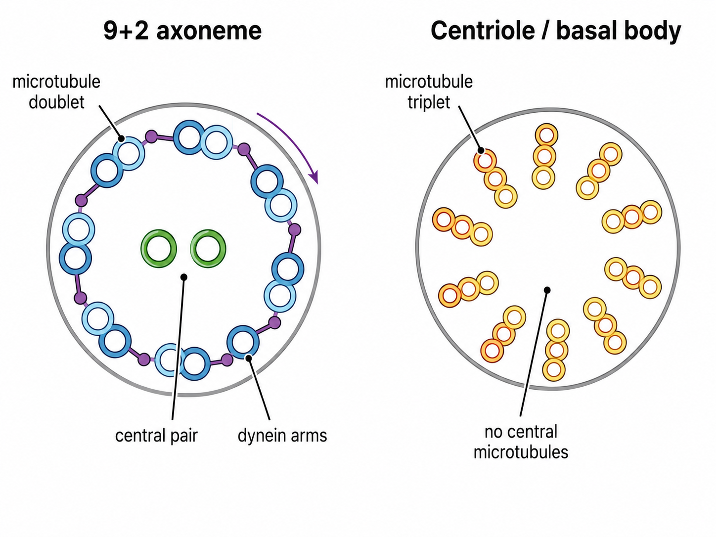
Must knowCilia and flagella are membrane projections with a core scaffold, the axoneme. Motile cilia/flagella have the "9+2" arrangement: nine outer doublet microtubules surrounding a central pair of singlets.
Know the logicThe motor is axonemal dynein, which projects as dynein arms from one doublet toward the next. Dynein walks toward the minus end, but because the doublets are crosslinked and anchored at the base, the sliding force is converted into bending — the beat of the cilium or flagellum.
Cilia vs. Flagella
Must know| Feature | Cilia | Flagella |
|---|---|---|
| Length / number | Short, many | Long, few (1 in sperm) |
| Beat | Coordinated, oar-like | Undulating wave |
| Examples | Respiratory & oviduct epithelium, ependyma | Sperm |
Functions of cilia: respiratory cilia sweep mucus (mucociliary escalator); oviduct cilia move the egg; ependymal cilia circulate CSF; nodal cilia establish left-right body asymmetry in development.
Eukaryotic vs. Prokaryotic Flagella
Must knowThe eukaryotic flagellum is the 9+2 microtubule axoneme, driven by axonemal dynein using ATP, and moves by bending. The prokaryotic flagellum is built from flagellin (not microtubules), powered by the proton-motive force (not ATP), and rotates like a rigid propeller.
Primary (Non-Motile) Cilia
Must knowPrimary cilia are sensory organelles on most non-dividing cells with a "9+0" arrangement (nine doublets, no central pair). They don't beat; they act as antennae sensing chemical gradients, flow, and signals (e.g., Hedgehog signaling).
Kartagener Syndrome (Primary Ciliary Dyskinesia)
Passage-levelA dynein arm defect — cilia/flagella form normally (9+2 present) but are immotile. Consequences: chronic respiratory infections (mucus not cleared), male infertility (sperm can't swim), and situs inversus (nodal cilia fail, randomizing left-right asymmetry; ~half have mirror-image organs). Triad: bronchiectasis + situs inversus + infertility.
Quick check: A Kartagener patient has sperm with normal 9+2 axonemes on EM but immotile. What is defective, and why doesn't 9+2 architecture guarantee motility?
Answer: The dynein arms are absent/nonfunctional. The 9+2 arrangement is the scaffold, but axonemal dynein is the motor that converts ATP into sliding and bending. Structure without the motor = no movement.
Centrioles and Microtubule-Organizing Centers
Centriole Structure
Must knowCentrioles are cylindrical, built from nine triplets of microtubules in a ring — a "9+0" triplet organization (no central tubules). Two centrioles at right angles, surrounded by pericentriolar material (PCM), form a centrosome, the primary microtubule-organizing center (MTOC) in animal cells.
Function as the MTOC
Must knowThe centrosome nucleates microtubules from PCM ring complexes of γ-tubulin; their minus ends anchor at the MTOC and plus ends extend outward (the radial, aster-like array). The centrosome duplicates once per cycle (S phase); at mitosis the two centrosomes separate to form the two spindle poles. Errors in centrosome number cause multipolar spindles and chromosomal instability (cancer).
Basal Body
Must knowThe basal body anchoring each cilium/flagellum is structurally identical to a centriole (9+0 triplet). A centriole migrates to the surface and becomes a basal body, from which the axoneme grows.
Must-memorize "9+X" chart:
| Structure | Arrangement | Notes |
|---|---|---|
| Motile cilia / flagella | 9+2 doublets | central pair; axonemal dynein drives movement |
| Primary cilia | 9+0 doublets | no central pair; non-motile, sensory |
| Centriole / Basal body | 9 triplets (9+0) | no center tubules; nucleates MTs or anchors axoneme |
Quick check
Plant cells lack centrioles yet still form a mitotic spindle and divide. What does this tell you about centrioles and spindle formation?
Answer: Centrioles are not required for spindle formation. Plant cells use other MTOC structures to nucleate the spindle; the fundamental requirement is γ-tubulin–containing PCM, not the centriole. Centrioles are needed for basal body/cilia formation, not mitosis.
Common Confusions & Tricks
1. 9+2 vs. 9+0. Motile cilia/flagella = 9+2 (central pair = movement). Centrioles/basal bodies = 9 triplets, 0 central (organize, don't move). Primary cilia = 9+0 doublets (doublets, not triplets; sensory).
2. Kinesin vs. dynein directions. Kinesin → plus end (cell periphery; plus ends point outward). Dynein → minus end (cell center). For cilia, axonemal dynein still moves toward the minus end, but crosslinked doublets bend instead of sliding.
3. IFs do NOT use nucleotide hydrolysis. Actin uses ATP; tubulin uses GTP; IFs have no polarity and no NTPase activity — don't say they "use ATP."
4. Kartagener triad — all three. Bronchiectasis + situs inversus + infertility. Students forget situs inversus, which the MCAT loves. Logic: nodal cilia are motile yet 9+0 and beat rotationally to drive leftward flow setting left-right asymmetry; when they fail, organ placement is random (~50% situs inversus).
5. Taxol vs. colchicine — opposite mechanisms, same outcome. A drug that "stabilizes" or "prevents depolymerization" acts like taxol; "destabilizes/prevents assembly" is colchicine. Both arrest mitosis.
6. Contractile ring uses microfilaments, not microtubules. Cytokinesis → contractile ring → actin + myosin II. Mitosis → spindle → microtubules. "Mitosis completes but cytokinesis fails" = an actin/microfilament defect.
7. Centrioles do NOT directly form spindle fibers. The PCM nucleates via γ-tubulin; centrioles organize the PCM. This is why plant cells (no centrioles) still form spindles.
8. Diameter order. Microfilaments (7 nm) < intermediate filaments (10 nm) < microtubules (25 nm). "7, 10, 25 — actin, IF, tubulin."
Key Takeaways
The Three Cytoskeletal Systems at a Glance
| Property | Microfilaments | Microtubules | Intermediate Filaments |
|---|---|---|---|
| Diameter | ~7 nm | ~25 nm | ~10 nm |
| Monomer | G-actin | α/β-tubulin dimer | Various (keratin, vimentin, desmin, neurofilaments, lamins) |
| Nucleotide | ATP | GTP | None |
| Polarity | Yes (+ barbed, − pointed) | Yes (+ fast, − slow) | No |
| Motor protein | Myosin | Kinesin (+), Dynein (−) | None |
| Primary role | Contraction, cleavage, motility | Transport, spindle, cilia | Structural support |
| Stability | Moderately dynamic | Highly dynamic (dynamic instability) | Most stable |
High-Yield Facts
- Contractile ring (actin + myosin II) drives cytokinesis; cytochalasin → binucleate cells.
- Treadmilling: actin adds at barbed (+) end, leaves pointed (−) end at equal rates.
- Dynamic instability: GTP cap loss → catastrophe; restored → rescue.
- Kinesin = anterograde (+ end, periphery); dynein = retrograde (− end, center).
- 9+2 doublets + axonemal dynein = motile cilia/flagella; 9+0 doublets = primary cilia (sensory); 9 triplets = centrioles/basal bodies.
- Kartagener = dynein arm defect → bronchiectasis + infertility + situs inversus.
- Centrosome = 2 centrioles + PCM; PCM nucleates microtubules via γ-tubulin.
- Taxol stabilizes (arrests anaphase); colchicine/vinblastine destabilize (block spindle assembly) — both arrest mitosis.
- Nuclear lamins (IFs) form the nuclear lamina; lamin A mutations → progeria.
- Basal body ≡ centriole (9 triplet); centrioles can differentiate into basal bodies.
- Plant cells lack centrioles but form spindles — centrioles are required for cilia/flagella, not mitosis.