The plasma membrane is heavily tested because it sits at the intersection of chemistry, physics, and biology. Every cell function — signaling, energy production, waste removal — depends on the membrane's ability to separate the cell from its environment while communicating with it.
Priority labels: Must know = cold; Know the logic = mechanism not names; Passage-level = recognize, don't memorize; Optional = skippable.
General Function in Cell Containment
Must knowThe plasma membrane defines the cell as a discrete unit and establishes selective permeability: it allows certain molecules in or out while excluding others. This lets the cell maintain internal conditions (ion concentrations, pH, macromolecule pools) far from equilibrium with the outside — a prerequisite for life. It also serves as a platform for signaling, adhesion, and identity (via surface proteins and carbohydrates).
Quick check: Why must the plasma membrane be selectively permeable rather than simply impermeable? Answer: A completely impermeable membrane would prevent the cell from taking in nutrients, exporting waste, or responding to signals — all essential life functions.
Composition of Membranes
Lipid Components
Must knowThe core is a phospholipid bilayer — two sheets of phospholipids arranged tail-to-tail. The driver is amphipathicity: each phospholipid has a hydrophilic head and two hydrophobic fatty acid tails. In water, thermodynamics buries the tails away from water while exposing the heads, making the bilayer the energetically favorable result.
A phospholipid = a glycerol backbone + two fatty acid tails (esterified at C1, C2) + a phosphate at C3 linked to a polar head group. The head group identity defines the class (phosphatidylcholine, -serine, etc.); "phosphatid-" just refers to this glycerophospholipid backbone. Passage-level you don't need to memorize individual head groups — just know that phosphatidylserine is normally on the inner leaflet and flips outward during apoptosis.
Sphingolipids are built on a sphingosine backbone rather than glycerol (e.g., sphingomyelin). Glycolipids (sugar head groups) sit exclusively on the extracellular face and contribute to the glycocalyx, the carbohydrate coat involved in cell recognition.
Cholesterol (a steroid) intercalates between phospholipid tails with its rigid planar ring. Its classic role is a fluidity buffer:
- At high temperature: restrains movement → decreases fluidity
- At low temperature: prevents tight packing → increases fluidity (prevents solidifying)
Waxes are esters of long-chain fatty acids with long-chain alcohols (not glycerol). Extremely hydrophobic; not membrane components but appear as protective barriers (plant cuticles, earwax).
Quick check: A researcher replaces all saturated fatty acids in a membrane with unsaturated fatty acids. What happens to fluidity and why? Answer: Fluidity increases. Unsaturated fatty acids contain double bonds that create kinks in the tails, preventing tight packing.
Protein Components
Must knowProteins make up roughly half of membrane mass and carry out most of its specific functions. Two major classes:
- Integral (intrinsic) proteins: embedded in the bilayer; many span it entirely (transmembrane proteins). They interact with the hydrophobic core via nonpolar side chains and require detergents to remove. Transmembrane segments are typically hydrophobic α-helices (or β-barrels in bacterial outer membranes).
- Peripheral (extrinsic) proteins: surface-associated (electrostatic interactions with head groups or integral proteins); removable by high salt or pH change without disrupting the bilayer.
A third group, lipid-anchored proteins (e.g., GPI-anchored), are covalently tethered to lipids. Membrane proteins serve as channels, transporters, receptors, enzymes, and anchors.
Quick check: A protein is solubilized only when the membrane is treated with a detergent such as SDS. Is it integral or peripheral? Answer: Integral — detergent disruption of the hydrophobic core is required.
Fluid Mosaic Model
Must knowThe fluid mosaic model is the accepted framework. Key ideas:
- The membrane is a two-dimensional fluid — lipids and many proteins diffuse laterally.
- Proteins are embedded in or attached to the bilayer (not sitting on a static sheet).
- The membrane is asymmetric: inner and outer leaflets differ in lipid and protein composition.
Lateral diffusion is fast. Flip-flop (transverse diffusion across leaflets) is rare and very slow without help, because moving the hydrophilic head through the hydrophobic core is thermodynamically costly; flippases catalyze it. Lipid rafts are cholesterol/sphingomyelin-rich microdomains thought to concentrate signaling molecules.
Quick check: Why is flip-flop (transverse diffusion) thermodynamically unfavorable without enzyme assistance? Answer: The hydrophilic phospholipid head group must pass through the hydrophobic interior of the bilayer — this has a very high energy barrier.
Membrane Dynamics
Must knowFluidity is the ease with which lipids and proteins move within the membrane. The MCAT asks you to predict how composition changes affect it — use the "kinks and space" model: anything that creates space between tails increases fluidity.
- Unsaturated tails → increase fluidity (kinks prevent packing)
- Shorter tails → increase fluidity (less van der Waals contact)
- Higher temperature → increases fluidity
- Cholesterol → buffers (decreases at high T, increases at low T)
Quick check: Deep-sea organisms living in cold environments tend to have membranes with a higher proportion of unsaturated fatty acids. Why is this adaptive? Answer: At cold temperatures, increased unsaturation keeps the membrane fluid (prevents solidification), maintaining function.
Solute Transport Across Membranes
Thermodynamic Considerations
Must knowPassive transport is governed by the electrochemical gradient — the concentration gradient plus, for charged solutes, the electrical gradient (membrane potential). Movement down the gradient is spontaneous (); active transport moves a solute against it () and must be coupled to an energy source. (Quantitative Nernst/Goldman treatment belongs to the electrochemistry/neuro guides — here the concept is what matters.)
Osmosis
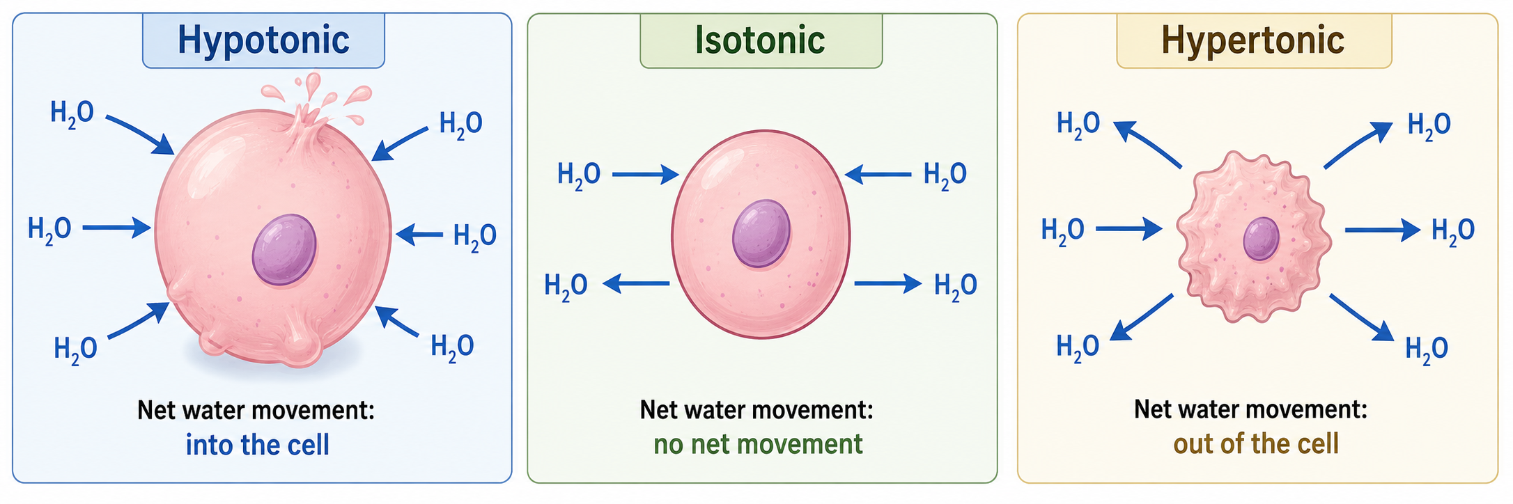
Must knowOsmosis is the net movement of water across a semipermeable membrane from lower solute concentration (higher water) to higher solute concentration (lower water) — water moves down its own gradient.
- Isotonic: equal solute inside/outside → no net water movement
- Hypotonic: lower solute outside → water enters → cell swells (lysis in animal cells; turgor in plants)
- Hypertonic: higher solute outside → water leaves → cell shrinks (crenation; plasmolysis in plants)
Colligative Properties and Osmotic Pressure
Must knowColligative properties depend on the number of dissolved particles, not their identity (vapor pressure lowering, boiling point elevation, freezing point depression, and osmotic pressure).
Osmotic pressure () is the pressure needed to prevent net osmotic flow:
where = van 't Hoff factor (particles per formula unit; glucose, NaCl), = molarity, = 0.08206 L·atm/(mol·K), = temperature (K).
Worked Example: Osmotic pressure of 0.15 M NaCl at 37°C (310 K)? NaCl dissociates, so :
This matches blood plasma (~7.6–7.7 atm), why 0.15 M NaCl is isotonic saline — a famous benchmark.
Passive Transport
Must knowPassive transport moves solutes down their electrochemical gradient with no cellular energy. Two subtypes:
- Simple diffusion: direct passage through the bilayer; limited to small nonpolar molecules (O₂, CO₂), small uncharged polar molecules (H₂O, urea, glycerol), and lipid-soluble molecules.
- Facilitated diffusion: uses a channel or carrier protein to move polar/charged molecules down their gradient — still no ATP. Examples: GLUT (glucose), aquaporins (water).
Simple diffusion rate follows Fick's law (): flux rises with the gradient and area.
Quick check: O₂ crosses the membrane by simple diffusion, but glucose requires GLUT transporters. What property of glucose makes simple diffusion insufficient? Answer: Glucose is a large, polar, hydrophilic molecule — it cannot partition into the hydrophobic membrane core.
Active Transport
Must knowActive transport moves solutes against their electrochemical gradient, requiring energy.
- Primary active transport: directly uses ATP hydrolysis (Na⁺/K⁺-ATPase, Ca²⁺-ATPase).
- Secondary active transport: uses a gradient set up by primary transport. Symport = two solutes same direction (SGLT: Na⁺ in drags glucose in); antiport = opposite directions (Na⁺/Ca²⁺ exchanger).
Sodium/Potassium Pump (Na⁺/K⁺-ATPase)
Must knowThe single most important transport protein on the MCAT.
- Moves 3 Na⁺ out and 2 K⁺ in per ATP
- Electrogenic: net 1 positive charge out per cycle → contributes to the negative resting potential
- Maintains the Na⁺ and K⁺ gradients that power secondary transport and action potentials
- Inhibited by ouabain and cardiac glycosides (digoxin), used in heart failure
It cycles between E1 (high Na⁺ affinity) and E2 (high K⁺ affinity) conformations driven by phosphorylation/dephosphorylation.
Quick check: A patient takes a drug that blocks the Na⁺/K⁺-ATPase. Predict what happens to intracellular [Na⁺] and [K⁺]. Answer: Intracellular [Na⁺] rises (pump no longer exports it) and intracellular [K⁺] falls (pump no longer imports it).
Membrane Channels
Must knowIon channels are integral proteins forming aqueous pores that let ions flow rapidly down their electrochemical gradients. Two key properties:
- Selectivity: channels admit specific ions based on pore size and selectivity-filter chemistry (the K⁺ channel selects K⁺ over the smaller Na⁺).
- Gating: channels open in response to a trigger — voltage-gated (action potentials), ligand-gated (nicotinic ACh receptor), or mechanically gated (hair cells).
Aquaporins are water channels explaining why water crosses some membranes far faster than diffusion predicts; AQP2 in the kidney collecting duct is regulated by ADH (vasopressin) — a favorite physiology connection.
Quick check: Why can't ions pass through aquaporins along with water? Answer: The aquaporin channel is engineered to exclude ions — it strips water molecules of their hydration shell one by one as they pass through, and the positive charge at the channel's center repels protons (H⁺).
Membrane Potential
Know the logicThe membrane potential () is the electrical difference across the membrane (inside minus outside); at rest in a typical animal cell mV (inside negative).
It arises because (1) the Na⁺/K⁺-ATPase builds steep gradients (high K⁺ in, high Na⁺ out), (2) at rest the membrane is far more permeable to K⁺ (open leak channels), and (3) K⁺ diffuses out, leaving the inside negative until the electrical pull back in balances the concentration push out. The resting potential (−70 mV) sits near the K⁺ equilibrium potential (−90 mV), pulled slightly positive by small Na⁺/Cl⁻ permeabilities. At MCAT level this is qualitative.
Quick check: Why is the resting membrane potential (−70 mV) closer to the K⁺ equilibrium potential (−90 mV) than to the Na⁺ equilibrium potential (~+60 mV)? Answer: At rest the membrane is far more permeable to K⁺ than to Na⁺ (open leak K⁺ channels), so the resting potential is dominated by K⁺ and sits near its equilibrium value, pulled only slightly positive by the small Na⁺ leak.
Membrane Receptors
Must knowMembrane receptors let the cell sense extracellular signals without admitting them (usually large, polar, or charged molecules). Ligand binding triggers a conformational change that starts an intracellular response (signal transduction). Major classes:
- Ion channel-linked (ionotropic): ligand binding opens an ion channel (nicotinic ACh receptor).
- G protein-coupled receptors (GPCRs): 7 transmembrane helices; activate G proteins → second messengers (cAMP, IP₃/DAG); e.g., β-adrenergic receptor.
- Receptor tyrosine kinases (RTKs): ligand → dimerization → autophosphorylation → cascade; e.g., insulin receptor.
- Intracellular receptors (not membrane receptors): for lipid-soluble ligands (steroids, thyroid hormone) that cross the membrane and regulate genes; e.g., estrogen receptor.
The MCAT contrasts these to test which signaling molecules can cross the membrane.
Quick check: Epinephrine is a water-soluble (catecholamine) hormone. Cortisol is a steroid hormone. Which one uses a membrane receptor and which one uses an intracellular receptor? Answer: Epinephrine (polar, cannot cross membrane) uses a GPCR membrane receptor. Cortisol (steroid, nonpolar, membrane-permeable) uses an intracellular receptor.
Exocytosis and Endocytosis
Must knowThese move large molecules or particles via vesicles (bulk transport).
Exocytosis fuses an intracellular vesicle with the plasma membrane, releasing contents outside (neurotransmitter release, insulin secretion). Regulated exocytosis is triggered by a Ca²⁺ rise; constitutive exocytosis is continuous.
Endocytosis invaginates the membrane to engulf material into a vesicle:
- Phagocytosis ("cell eating"): engulfs large particles → phagosome → fuses with lysosome → degradation (macrophages, neutrophils).
- Pinocytosis ("cell drinking"): non-specific uptake of fluid and dissolved solutes.
- Receptor-mediated endocytosis (RME): selective; ligands bind receptors in clathrin-coated pits → vesicle → endosome → recycled or sent to lysosome. Archetype: LDL receptor (defective in familial hypercholesterolemia).
Quick check: A macrophage engulfs a bacterium. Trace the path from phagocytosis to degradation. Answer: Pseudopods extend around the bacterium → phagosome forms → lysosome fuses with phagosome → phagolysosome → lysosomal hydrolases (active at low pH) degrade bacterium → products released.
Intercellular Junctions
Must knowThree junction types are tested:
- Tight junctions (zonula occludens): a seal between adjacent epithelial cells (claudins, occludins) that blocks paracellular transport and maintains cell polarity. Connections: blood-brain barrier, gut, kidney tubules.
- Gap junctions: direct cytoplasmic channels formed by connexons (from connexin), allowing passage of ions and small molecules (<~1,000 Da). Enable electrical coupling — key in cardiac muscle for synchronized contraction.
- Desmosomes (maculae adherentes): strong anchor junctions linking intermediate filaments (keratin) of adjacent cells via cadherins; purely structural, resisting mechanical stress. Optional hemidesmosomes anchor cells to the extracellular matrix; pemphigus vulgaris attacks desmosomal cadherins → blistering.
| Junction | Proteins | Function |
|---|---|---|
| Tight | Claudins, Occludins | Seal; block paracellular transport |
| Gap | Connexins (connexons) | Direct cell–cell communication |
| Desmosome | Cadherins | Mechanical adhesion via intermediate filaments |
Quick check: The heart contracts as a single unit even though individual cardiomyocytes are separate cells. Which junction type is responsible? Answer: Gap junctions — they electrically couple cardiomyocytes so action potentials spread from cell to cell.
Common Confusions & Tricks
1. Osmosis direction: think about WATER, not solute. Water moves from low solute (high water) to high solute (low water). Mnemonic: water runs away from salt.
2. Hypo vs. hyper relative to what? Hypotonic = outside has fewer solutes → water enters → cell swells. Hypertonic = outside has more → water leaves → cell shrinks. Always ask "relative to what?"
3. The Na⁺/K⁺ pump stoichiometry: 3 out, 2 in. Students reverse this. Remember it's also electrogenic (net positive charge leaves).
4. Active vs. secondary active transport. Secondary active transport moves something against its gradient but doesn't directly use ATP. Inhibit the Na⁺/K⁺-ATPase → intestinal glucose absorption stops, because SGLT depends on the Na⁺ gradient. A classic reasoning chain.
5. Facilitated diffusion is still passive. GLUT, aquaporins, and ion channels don't consume ATP. "Facilitated" just means protein-assisted; the driver is still the gradient.
6. Integral vs. peripheral proteins — detergent test. Detergent required → integral. Salt/pH change sufficient → peripheral.
7. Cholesterol as a fluidity BUFFER, not just an inhibitor. At physiological temperature it decreases fluidity; at low temperature it prevents gelling. The MCAT tests both directions.
8. Tight junctions seal; desmosomes hold; gap junctions talk.
9. Endocytosis vs. exocytosis balance. Exocytosis adds membrane; endocytosis removes it. The two stay balanced at steady state.
10. Receptor-mediated endocytosis requires a LIGAND binding first. Don't confuse it with pinocytosis (nonspecific). LDL receptor → coated pit → endosome is the archetype.
11. Steroids use intracellular receptors; peptide hormones use membrane receptors. Steroids are lipid-soluble (cross freely); peptide hormones are polar (can't cross).
Key Equations
| Equation | Variables / Notes | When to Use |
|---|---|---|
| : osmotic pressure (atm); : van 't Hoff factor; : molarity; : 0.08206 L·atm/mol·K; : temperature (K) | Osmotic pressure; comparing osmolarity | |
| : flux; : diffusion coefficient; : area; : gradient | Rate/direction of simple diffusion (Fick's first law) | |
| : boiling point elevation; : ebullioscopic constant; : molality | Colligative — boiling point elevation | |
| : freezing point depression; : cryoscopic constant; : molality | Colligative — freezing point depression |