Introduction: Why Tissues Matter on the MCAT
Multicellular life depends on specialization: groups of cells with similar structure and origin cooperate to perform a specific function — a tissue. The four primary types are epithelial, connective, muscle, and nervous; this guide covers the two most heavily tested, epithelial and connective. The MCAT's recurring move is asking why a tissue's structure matches its function.
Priority labels: Must know = cold; Know the logic = mechanism not names; Passage-level = recognize, don't memorize; Optional = skippable.
Epithelial Cells
The Core Idea: Barriers and Interfaces
Must knowEpithelial tissue lines every body surface that meets the outside world or an internal cavity — skin, gut lumen, airways, blood vessels, glands. Its defining feature is polarity: an apical surface (facing the lumen/exterior) and a basal surface (facing underlying tissue), with the basal side resting on a non-cellular basement membrane (basal lamina). Polarity allows directional transport — e.g., intestinal cells absorb nutrients apically and export them basally into the blood.
Epithelial cells are tightly packed, have little extracellular matrix, and are generally avascular, receiving nutrients by diffusion from capillaries in the connective tissue below.
Classification: Shape × Layering
Must knowTwo words — shape and number of layers — describe any epithelium, and the combination predicts function.
Shape: squamous (flat, scale-like) → cuboidal (cube-shaped) → columnar (taller than wide). Layering:
- Simple — single layer; every cell touches the basement membrane. Optimized for diffusion/transport.
- Stratified — multiple stacked layers; only the bottom layer touches the basement membrane. Optimized for protection. Named for the apical layer's shape (skin = stratified squamous even though basal cells aren't flat).
- Pseudostratified — looks layered but is actually simple (all cells touch the basement membrane); nuclei at different heights create a false impression of layers. Classic: pseudostratified ciliated columnar epithelium of the trachea/bronchi.
Transitional epithelium (urothelium) lines the urinary tract and stretches as the bladder fills — a structure-matches-function favorite; simple squamous lining vessels = endothelium, lining body cavities = mesothelium; stratified squamous appears where abrasion is likely (skin, mouth, esophagus).
Apical Specializations
Must know- Microvilli: finger-like membrane projections that amplify surface area for absorption (intestinal "brush border," kidney proximal tubule). Non-motile.
- Cilia: longer, motile projections (9+2 microtubules powered by dynein) that propel material — mucus clearance in airways, ovum transport in the fallopian tube. (Primary cilia, 9+0, are non-motile sensory structures.)
- Stereocilia: elongated microvilli, not true cilia; inner ear and epididymis.
Cell Junctions: Holding the Barrier Together
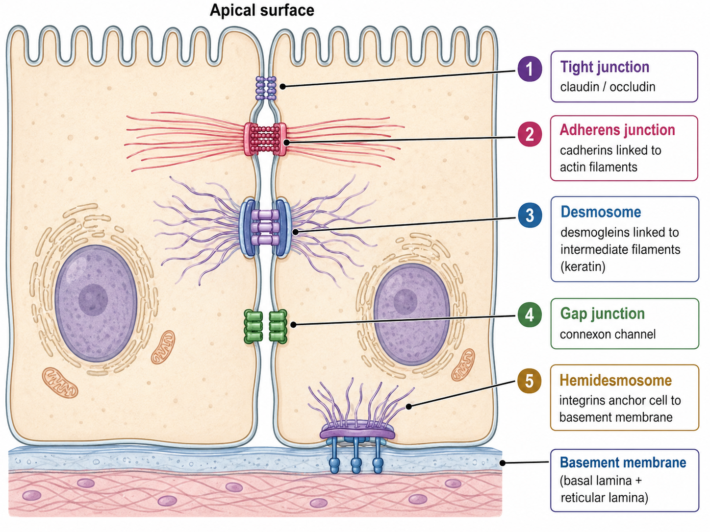
Must knowKnow these by function and intracellular anchor:
- Tight junctions (zonula occludens): fuse adjacent membranes (claudin, occludin) to block paracellular leakage between cells and enforce polarity. Key in the blood-brain barrier and gut.
- Adherens junctions: belt of E-cadherin linked to actin; mechanical cohesion. Hook: loss of E-cadherin marks epithelial-to-mesenchymal transition in metastasis.
- Desmosomes: spot welds of cadherins (desmogleins) linked to intermediate filaments (keratin); resist shear. Hook: pemphigus vulgaris = autoantibodies to desmogleins → blistering.
- Gap junctions: connexin channels directly coupling cytoplasms (ions, small molecules); electrical/metabolic coupling, crucial in cardiac and smooth muscle.
- Hemidesmosomes: anchor the basal surface to the basement membrane via integrins linked to intermediate filaments.
Apical→basal order: Tight → Adherens → Desmosome → Gap (mnemonic TADG).
Glands: Epithelial Secretory Organs
Must knowGlands form from epithelial invaginations and specialize in secretion.
- Endocrine glands: no duct; secrete hormones directly into the bloodstream (thyroid, adrenal, pituitary, pancreatic islets).
- Exocrine glands: retain a duct; secrete onto a surface or into a cavity (sweat, salivary, pancreatic acini, sebaceous).
Exocrine secretion modes: merocrine/eccrine = exocytosis, no cell loss (most sweat glands); apocrine = apical cytoplasm buds off (mammary); holocrine = whole cell disintegrates into secretion (sebaceous).
Quick check: A slide shows a single layer of cells, all touching the basement membrane, nuclei at varying heights, with cilia on the apical surface. What epithelium is this, and where is it found?
Answer: Pseudostratified ciliated columnar epithelium, lining the trachea and bronchi. Varying nuclear heights mimic layering, but every cell contacts the basement membrane, so it is simple.
Connective Tissue Cells
The Core Idea: Support, Structure, and Communication
Must knowWhere epithelium is mostly cells, connective tissue is mostly extracellular matrix (ECM) — a scaffold of proteins and polysaccharides secreted by dispersed cells. Connective tissues are generally vascular (cartilage is the exception). All the variety (soft adipose to rigid bone) comes from differences in ECM composition produced by a small cast of cell types.
The Extracellular Matrix
Must know- Collagen: most abundant protein in the body; triple-helical fibers giving tensile strength. Vitamin C is required for stable collagen, so deficiency causes scurvy (fragile vessels, bleeding gums, poor healing). (Collagen type-by-type and the hydroxylation pathway are out of scope.)
- Elastin: elastic fibers that stretch and recoil (elasticity); abundant in large arteries, lung, skin. Collagen prevents over-stretch; elastin enables recoil.
- Reticular fibers (type III collagen): fine mesh forming the stroma of lymph nodes, spleen, marrow.
- Proteoglycans / GAGs: negatively charged, heavily hydrated polysaccharides that resist compression — why cartilage cushions joints. Fibronectin links cells to the matrix via integrins.
Connective Tissue Cell Types
Must knowKnow each cell's product/role:
- Fibroblasts: most common; synthesize ECM (collagen, elastin, fibronectin, proteoglycans); drive wound healing. Excess activity → fibrosis.
- Adipocytes: fat storage. White adipose = one large lipid droplet; stores energy, insulates, cushions, and acts as an endocrine organ (secretes leptin). Brown adipose (BAT) is mitochondria-rich and uses uncoupling protein 1 (UCP-1/thermogenin) to uncouple electron transport from ATP synthesis, dissipating the proton gradient as heat (non-shivering thermogenesis). MCAT hook: UCP-1 in the proton-gradient/ATP-synthesis context.
- Mast cells: immune cells with granules of histamine (and heparin); IgE-triggered degranulation drives allergic responses; antihistamines block histamine receptors.
- Macrophages: monocyte-derived phagocytes; also antigen presentation and cytokine secretion. Tissue-specific names (Kupffer cells in liver, microglia in brain) are passage-level.
- Plasma cells: terminally differentiated B cells; antibody factories with abundant rough ER.
Specialized Connective Tissues
Cartilage
Must knowCartilage is avascular, so nutrients reach chondrocytes (sitting in lacunae) only by diffusion — which is why it heals slowly. Chondroblasts produce matrix and mature into chondrocytes.
Know the logicHyaline (type II collagen + proteoglycans; articular surfaces, trachea, fetal skeleton — smooth load-bearing); elastic (+ elastin; ear, epiglottis — flexible); fibrocartilage (dense type I collagen; intervertebral discs, menisci — strongest, shock-absorbing).
Bone
Must knowBone ECM is mineralized: hydroxyapatite () on a type I collagen scaffold — mineral gives hardness, collagen gives toughness.
Know the four cell types and origins:
| Cell | Origin | Function |
|---|---|---|
| Osteoblasts | Mesenchymal | Secrete and mineralize bone matrix (osteoid) |
| Osteocytes | Trapped osteoblasts | Maintain bone; reside in lacunae, communicate via canaliculi; sense load |
| Osteoclasts | Monocyte/macrophage (hematopoietic) | Resorb bone via acid () and proteases |
| Osteoprogenitors | Mesenchymal | Osteoblast precursors |
Bone remodeling couples osteoclast resorption with osteoblast deposition, regulated by PTH (raises blood calcium via osteoclasts), calcitonin (lowers it), vitamin D, and mechanical loading.
Passage-levelCompact bone = osteons (Haversian systems), concentric lamellae around a Haversian canal; spongy bone = trabeculae aligned to stress lines, housing red marrow.
Blood
Must knowBlood is a connective tissue: cells (erythrocytes, leukocytes, platelets) in a fluid ECM, plasma (proteins, ions, nutrients, gases). It fits the definition — cells of mesodermal origin in a non-cellular matrix.
Types of Connective Tissue Proper
Must knowClassified by matrix density and organization:
- Loose (areolar): widely spaced fibers, abundant ground substance; houses fibroblasts, macrophages, mast cells; sits under epithelium (gut lamina propria) for immune surveillance.
- Dense regular: parallel collagen for unidirectional tensile strength — tendons (muscle→bone), ligaments (bone→bone).
- Dense irregular: randomly oriented collagen resisting multidirectional force — dermis, joint capsules.
- Adipose (sparse matrix, adipocyte-dominated) and reticular (type III mesh) are also connective tissue proper.
Quick check: A patient develops scurvy after months without vitamin C. Which cell type is most directly affected, which protein is deficient, and what happens in blood vessel walls?
Answer: Fibroblasts (the main collagen producers) — without vitamin C they cannot make stable collagen. Vessel walls rely on collagen for tensile strength, so deficiency causes fragile capillaries, easy bruising, and bleeding gums.
Common Confusions & Tricks
1. Pseudostratified ≠ stratified. Every cell in pseudostratified epithelium touches the basement membrane — it is fundamentally simple. Varying nuclear heights alone don't make it stratified.
2. Stratified naming follows the apical layer. Skin is stratified squamous even though basal cells aren't flat.
3. Microvilli vs. cilia — motility. Microvilli = non-motile, absorptive (brush border). Cilia = motile, propulsive. (Kartagener syndrome: immotile cilia → respiratory + fertility problems, since sperm flagella use the same dynein.)
4. Tight junctions seal; gap junctions communicate. Tight = block material between cells; gap = let ions/molecules pass between cells.
5. Desmosomes → intermediate filaments; adherens → actin. The intracellular anchor differs.
6. Osteoclasts are NOT osteoblast-derived. Osteoclasts = monocyte/macrophage (hematopoietic); osteoblasts/osteocytes/osteoprogenitors = mesenchymal.
7. Cartilage heals slowly because it is avascular — no blood vessels means no rapid repair response reaches it.
8. Brown fat ≠ white fat. White stores energy; brown burns it as heat via UCP-1. Multilocular, mitochondria-rich fat cells = brown adipose.
9. Blood is connective tissue — cells in a plasma ECM, mesodermal origin.
10. Endocrine vs. exocrine — the duct. Endocrine = no duct, into blood; exocrine = duct, onto a surface. The pancreas is both (acini exocrine; islets endocrine).
Key Takeaways
Epithelial Tissue
- Polarity: apical vs. basal (rests on basement membrane); enables directional transport. Cell-rich, avascular.
- Shape: squamous → cuboidal → columnar. Layering: simple (all touch BM) → pseudostratified (looks layered, all touch BM) → stratified (true layers, named by apical shape).
- Apical: microvilli = non-motile absorption; cilia = motile (9+2 dynein) propulsion; stereocilia = elongated microvilli.
- Junctions (apical→basal): Tight (seal) → Adherens (actin, E-cadherin) → Desmosome (intermediate filaments, desmogleins) → Gap (connexin, communication); Hemidesmosomes anchor basal surface to BM via integrins.
- Glands: endocrine (no duct, blood) vs. exocrine (duct, surface); secretion modes merocrine/apocrine/holocrine.
Connective Tissue
- ECM dominant: collagen (tension), elastin (recoil), proteoglycans (compression). Vitamin C → stable collagen → scurvy if deficient. Usually vascular except cartilage.
| Cell | What it does |
|---|---|
| Fibroblast | Synthesizes ECM; drives wound healing |
| Adipocyte | Stores fat (white) or generates heat via UCP-1 (brown) |
| Mast cell | Releases histamine; allergic responses |
| Macrophage | Phagocytosis; antigen presentation; cytokines |
| Plasma cell | Antibody secretion; differentiated B cell |
| Osteoblast | Bone matrix synthesis (mesenchymal) |
| Osteoclast | Bone resorption (monocyte/hematopoietic) |
| Osteocyte | Mature osteoblast; maintains bone; senses load |
| Chondroblast/chondrocyte | Cartilage matrix; avascular |
- Cartilage: hyaline (articular/trachea, type II) → elastic (ear, + elastin) → fibrocartilage (discs, type I, strongest).
- Bone: hydroxyapatite on collagen; compact = osteons; spongy = trabeculae aligned to stress.
- Blood = connective tissue (cells in plasma ECM).
- Connective tissue proper: loose (immune surveillance) → dense regular (tendons/ligaments) → dense irregular (dermis).