Overview: From One Cell to an Organism
Embryogenesis is a precisely orchestrated sequence of division, movement, signaling, and specialization in which cells progressively commit to specific fates. Think of it as a series of increasingly refined decisions: first "how many cells?", then "which germ layer?", then "what tissue?"
The MCAT tests the order of stages, the key events within each, the germ layer derivatives, and how genes and environment collaborate to build complexity.
Priority labels: Must know = cold; Know the logic = mechanism not names; Passage-level = recognize, don't memorize; Optional = skippable.
Fertilization
The Events of Fertilization
Must knowFertilization is the fusion of a haploid sperm and a haploid secondary oocyte to produce a diploid zygote. It typically occurs in the ampulla of the fallopian tube. The secondary oocyte is arrested in metaphase II and only completes meiosis II after a sperm penetrates it — so what gets fertilized is a secondary oocyte, not a mature egg.
The sperm passes through the corona radiata (surrounding follicular cells) and then the zona pellucida (glycoprotein coat). At the zona it undergoes the acrosomal reaction: the acrosome releases hydrolytic enzymes that digest through the zona pellucida, letting the sperm reach and fuse with the oocyte membrane.
Blocks to Polyspermy
Must knowIf more than one sperm fertilizes an egg (polyspermy), the cell is non-viable. Two mechanisms prevent this:
-
Fast block — On sperm-egg fusion, the oocyte membrane immediately depolarizes, transiently preventing additional fusion. Fast (seconds) but temporary.
-
Slow block (cortical reaction) — release triggers cortical granules to exocytose, hardening the zona pellucida into a fertilization membrane — a permanent physical barrier. Slower (minutes) but definitive.
Following fusion, the oocyte completes meiosis II, the two pronuclei fuse, and the diploid zygote begins cleavage.
Quick check: A student says, "The oocyte that gets fertilized has already completed meiosis." Is this correct?
Answer: No. A secondary oocyte is arrested in metaphase II and only completes meiosis II after fertilization is initiated. (Primary oocytes are arrested earlier, in prophase I.)
Cleavage
Rapid Division Without Growth
Must knowAfter fertilization, the zygote undergoes rapid mitotic divisions called cleavage. The defining feature: there is no cell growth between divisions — total cytoplasmic volume stays the same while the number of cells (blastomeres) increases, so each blastomere is smaller than the last. Because the nucleus stays full-sized, cleavage raises the nuclear-to-cytoplasmic ratio. The solid ball of cells produced is the morula.
Types of Cleavage
Know the logicTwo independent distinctions:
- Holoblastic (entire egg divides; little yolk — mammals, sea urchins) vs. meroblastic (only part divides; yolk-rich regions excluded — birds, reptiles, fish). Humans are holoblastic.
- Determinate (mosaic) cleavage: fate set early by segregated cytoplasmic determinants; an isolated blastomere forms only a partial structure (typical of protostomes). Indeterminate (regulative) cleavage: early blastomeres remain totipotent, so an isolated blastomere can form a complete embryo (deuterostomes, including mammals).
Twinning follows: monozygotic ("identical") twins arise when one zygote's still-totipotent cells split (earlier split → more separate membranes). Dizygotic ("fraternal") twins come from two eggs fertilized by two sperm — genetically just siblings.
Quick check: Why do blastomeres get progressively smaller during cleavage?
Answer: Because there is no growth phase between divisions — the original zygote's cytoplasm is partitioned into more cells without adding mass.
Blastula Formation
Forming the Blastocyst
Must knowAfter the morula reaches the uterus, fluid accumulates to form a cavity called the blastocoel. The structure is now a blastula (in mammals, a blastocyst). Two cell populations emerge:
| Cell population | Location | Fate |
|---|---|---|
| Inner cell mass (ICM) / embryoblast | Interior, one side | The embryo proper (all three germ layers) + some extraembryonic membranes |
| Trophoblast | Outer layer | Placenta and extraembryonic structures; does NOT become the embryo |
The ICM is pluripotent (can become any cell of the body, but not a whole organism) — this is why embryonic stem cells come from it. Truly totipotent cells exist earlier (2- to 4-cell stage).
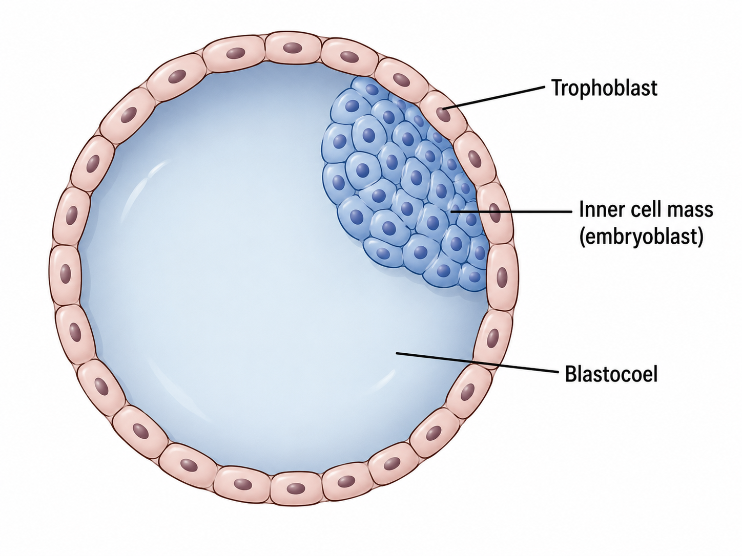
The blastocyst then undergoes implantation into the uterine endometrium (~day 6–10); the trophoblast invades and forms the placenta.
Quick check: A researcher isolates trophoblast cells and implants them into a surrogate. Could these cells form a complete embryo?
Answer: No. The trophoblast gives rise only to placental/extraembryonic structures. Only ICM cells have the pluripotency to form embryonic tissues.
Gastrulation
Rearranging the Blueprint
Must knowGastrulation reorganizes the single-layered blastula into three layers. This is the key stage for the MCAT because it establishes the three primary germ layers — ectoderm, mesoderm, endoderm.
In humans (and other amniotes), surface cells of the embryonic disc migrate toward a midline primitive streak, which marks the future anterior-posterior axis and is the site where cells ingress to form the internal layers.
First Cell Movements and Germ Layer Formation
Know the logicCells of the epiblast migrate through the primitive streak: the first wave displaces the hypoblast to form endoderm (innermost); a second wave spreads between to form mesoderm (middle); cells that stay on the surface become ectoderm (outermost). Net result: ecto → outside, meso → middle, endo → inside.
In sea urchins and frogs (common passage organisms), gastrulation involves the blastopore — an opening through which cells invaginate to form the archenteron (primitive gut). Must know blastopore fate: in deuterostomes (echinoderms, chordates, humans) the blastopore becomes the anus; in protostomes it becomes the mouth.
Quick check: After gastrulation, label the three layers from outside to inside.
Answer: Ectoderm (outside) → Mesoderm (middle) → Endoderm (inside).
Primary Germ Layer Derivatives
The Most-Tested Content in This Unit
Must knowGerm layer derivatives appear constantly, often clinically ("this tumor arises from mesoderm-derived tissue — what is it?").
Ectoderm = skin + brain (plus neural crest). Epidermis (hair, nails, glands); entire CNS (via neurulation); PNS (via neural crest); lens/cornea/retina; inner ear; adrenal medulla and anterior pituitary; tooth enamel.
Mesoderm = muscle, bone, blood, and deep organs. All muscle; bone, cartilage, connective tissue (incl. dermis); circulatory system (heart, vessels, blood cells); kidneys; gonads and reproductive ducts; adrenal cortex; spleen; serosal linings.
OptionalSub-framework: mesoderm organizes medial → lateral into paraxial (somites → vertebrae, skeletal muscle, dermis), intermediate (urogenital system), and lateral plate (circulatory system, serosal linings, limb connective tissue).
Endoderm = epithelial linings of tubes and glands. Epithelium of the GI tract and respiratory tract; liver and pancreas (gut outgrowths); thyroid, parathyroid, thymus; lining of bladder and urethra. (Note: posterior pituitary is neural ectoderm, anterior pituitary is oral ectoderm — neither is endoderm.)
Quick check: A patient develops a tumor of the adrenal cortex (secretes cortisol/aldosterone). What germ layer is the cortex from? What about the medulla?
Answer: Cortex = mesoderm; medulla = neural crest cells (ectoderm). The adrenal gland has a dual embryological origin — one of the most tested germ layer facts.
Neurulation
From Flat Sheet to Neural Tube
Know the logicNeurulation forms the neural tube (precursor to brain and spinal cord) from dorsal ectoderm. It is induced by the notochord (a mesodermal rod that later persists as the nucleus pulposus of intervertebral discs).
Sequence: notochord signals induce the ectoderm to thicken into the neural plate → its edges rise as neural folds → the folds fuse at the midline, pinching off the neural tube (brain anterior, spinal cord posterior), with surface ectoderm closing over the top.
Neural Tube Defects
Must knowFailure to close causes neural tube defects (NTDs): anencephaly (anterior neuropore fails; fatal) and spina bifida (posterior neuropore fails). Clinical hook: adequate folate (folic acid) before and during early pregnancy markedly reduces NTD risk.
Quick check: The notochord induces the neural plate. What does the notochord become in adult humans?
Answer: Its remnant persists as the nucleus pulposus — the gelatinous center of each intervertebral disc (herniation = "slipped disc").
Neural Crest Cells
The "Fourth Germ Layer"
Must knowAs the neural tube closes, cells at the junction between the neural tube and surface ectoderm delaminate and migrate throughout the embryo. They arise from ectoderm but travel far, giving a remarkably broad set of derivatives — hence "fourth germ layer."
Key derivatives: peripheral nervous system (sensory/autonomic ganglia), Schwann cells, adrenal medulla (catecholamines), melanocytes, craniofacial bone/cartilage. Reference (lower-yield): C cells of thyroid, odontoblasts, aorticopulmonary septum.
The pattern: neural crest → sensory/autonomic neurons, pigment cells, craniofacial structures.
Quick check: A newborn has a pheochromocytoma (catecholamine-secreting tumor of the adrenal medulla). From which germ layer are the cells of origin derived?
Answer: Neural crest cells, which arise from ectoderm. Even though the tumor is in the adrenal gland, the medullary cells are ectodermal in origin; the cortex, by contrast, is mesodermal.
Environment–Gene Interaction in Development
Induction and Competence
Know the logicDevelopment is a dialogue between cells and environment. Induction is the process by which one group of cells (the inducer) signals an adjacent group (the responding tissue) to change its fate. For this to work, the responding tissue must be competent — express the right receptors and machinery — and competence is often transient (a limited developmental window).
The Spemann–Mangold Organizer
Must knowSpemann and Mangold transplanted the dorsal lip of the blastopore from one salamander gastrula to another, inducing a second complete body axis. The transplanted cells acted as an organizer, instructing host cells to form neural/axial structures. The takeaways: a specific region can instruct adjacent fates via diffusible signals, and the responder must be competent. The vertebrate equivalent is the notochord inducing the neural plate.
Morphogens and Concentration Gradients
Know the logicA morphogen diffuses from a source and forms a concentration gradient; cells at different positions receive different concentrations and adopt different fates. The key idea is positional information — a cell "reads" its morphogen concentration like an address. (Specific morphogen identities and Hox genes are beyond MCAT scope here.)
Apoptosis in Development
Know the logicProgrammed cell death (apoptosis) is an essential sculpting tool — e.g., elimination of interdigital webbing to separate the digits, and pruning of excess neurons.
Stem Cell Potency
Must know| Term | Definition | Example |
|---|---|---|
| Totipotent | Any cell, including extraembryonic | Zygote; first 2–4 blastomeres |
| Pluripotent | Any cell of the embryo proper (not placenta) | ICM / embryonic stem cells |
| Multipotent | Multiple related cell types | Hematopoietic stem cells |
| Unipotent | One cell type | Spermatogonia |
Teratogens
Must knowTeratogens are environmental agents that disrupt development and cause birth defects, especially during organogenesis (~weeks 3–8). Classic examples: alcohol (fetal alcohol syndrome), certain drugs/toxins, and infections (e.g., rubella). The same agent produces different defects depending on when exposure occurs, because different tissues are competent and vulnerable at different windows — phenotype emerges from genotype and environment.
Quick check: Why does the timing of a teratogen exposure matter so much for the type of defect produced?
Answer: Because different organs form (and are most vulnerable) during specific windows, when their cells are competent to respond to signals. The same teratogen disrupts whichever structures are actively forming at that moment, producing different defects.
Common Confusions & Tricks
1. Adrenal cortex vs. medulla — the most-tested germ layer fact: Cortex = mesoderm (makes steroids). Medulla = neural crest/ectoderm (makes catecholamines). If the MCAT says "adrenal," ask which zone.
2. Anterior vs. posterior pituitary: Anterior (adenohypophysis) → oral ectoderm (Rathke's pouch); posterior (neurohypophysis) → neural ectoderm. Neither is endoderm.
3. Blastula ≠ blastomere: A blastomere is one cell from cleavage; the blastula/blastocyst is the whole fluid-filled embryo.
4. Trophoblast becomes placenta, NOT the embryo: The embryo comes only from the inner cell mass (ICM).
5. Protostome vs. deuterostome blastopore fate: In humans (deuterostomes): blastopore → anus. In protostomes: → mouth.
6. Secondary oocyte, not "egg," is what gets fertilized: Arrested in metaphase II; completes meiosis II only after sperm entry.
7. Fast vs. slow block: Fast (depolarization) = seconds, temporary. Slow (cortical reaction/zona hardening) = minutes, permanent.
8. Neural tube vs. neural crest: Neural tube → CNS. Neural crest → PNS, adrenal medulla, melanocytes, craniofacial structures.
9. Liver and pancreas are endoderm, not mesoderm: The functional parenchyma is an endodermal outgrowth of the gut tube (despite being "deep" in the body).
10. "If you see cleavage, think no growth": Cells get smaller; total mass constant, cell number increases.
Key Takeaways
Stage Sequence
Zygote → Cleavage (blastomeres, morula) → Blastula/blastocyst (ICM + trophoblast) → Gastrulation (3 germ layers via primitive streak) → Neurulation (neural plate → neural folds → neural tube) → Organogenesis
Fertilization Essentials
- Occurs in fallopian tube ampulla; oocyte in metaphase II
- Acrosomal reaction: sperm enzymes digest zona pellucida
- Fast block: depolarization (seconds, temporary); slow block: cortical granules harden zona (minutes, permanent)
Germ Layer Derivatives (Must Memorize)
| Germ Layer | Key Derivatives |
|---|---|
| Ectoderm | Epidermis, CNS, lens, cornea, anterior pituitary, adrenal medulla* |
| Mesoderm | Muscle, bone, connective tissue, heart, blood, kidneys, gonads, adrenal cortex |
| Endoderm | GI/respiratory epithelium, liver, pancreas, thyroid, parathyroid, thymus, bladder lining |
*via neural crest migration
Neural Crest Cell Derivatives
PNS, Schwann cells, adrenal medulla, melanocytes, craniofacial bone/cartilage (+ C cells of thyroid, aorticopulmonary septum)
Neurulation
Notochord (mesoderm) → induces neural plate → neural folds → neural tube (CNS). Failure to close → NTDs (anencephaly, spina bifida); prevented by folic acid.
Environment–Gene Interaction
- Induction: inducer signals responder to change fate; requires competence
- Spemann–Mangold organizer: dorsal lip of blastopore induces a second body axis
- Morphogens: concentration gradients give cells positional information
- Teratogens: agents (alcohol, drugs, infections) that disrupt development; effect depends on timing
- Apoptosis: sculpting tool (e.g., digit separation)
- Stem cell potency: totipotent (zygote) → pluripotent (ICM) → multipotent (adult stem cells)