A single fertilized cell gives rise to trillions of specialized cells. The MCAT tests how cells know what to become — the signaling, gene regulation, and physical mechanisms that guide a zygote through differentiation and even death. This guide runs from determination through senescence.
Priority labels: Must know = cold; Know the logic = mechanism not names; Passage-level = recognize, don't memorize; Optional = skippable.
Cell Specialization, Determination, and Differentiation
From Totipotency to Specialization
Must knowEarly embryonic cells have every option open; as development proceeds, options are progressively eliminated until each cell is committed. This narrows in two stages: determination then differentiation.
Determination is the internal commitment of a cell to a fate, before any visible change — it is heritable through divisions and not easily reversed. It is driven by changes in gene expression, triggered either by signals from neighbors or by inherited cytoplasmic determinants (fate-specifying mRNAs/proteins distributed unequally in the egg cytoplasm, so cleavage parcels different determinants into different blastomeres before any cell-cell signaling). Differentiation is the visible execution of that fate — the structural/functional changes that produce a muscle cell or neuron. Determination (invisible) precedes differentiation (visible).
Levels of Potency
Must knowCells exist on a hierarchy of developmental potential:
| Term | Definition | Example |
|---|---|---|
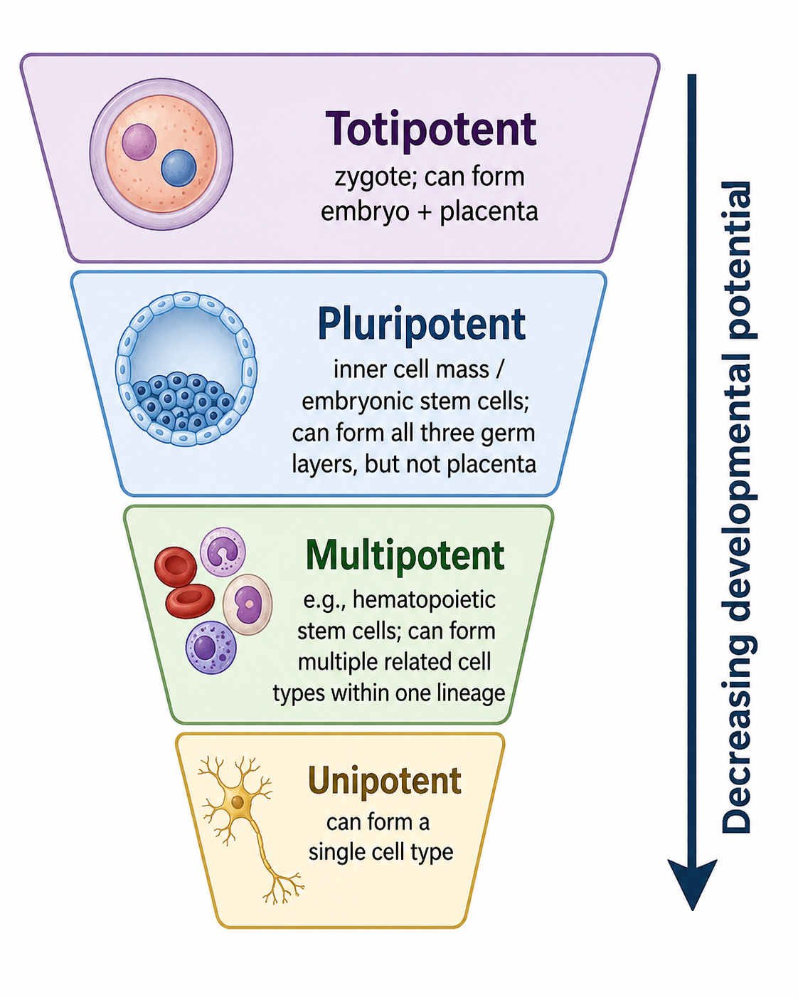
| Totipotent | Can form any cell type, including extraembryonic (placental) tissue | Zygote; cells through ~4-cell stage |
| Pluripotent | Can form any cell of the three germ layers, but NOT placenta | Inner cell mass (ICM) of blastocyst; embryonic stem cells |
| Multipotent | Can form multiple, but limited, cell types within a lineage | Hematopoietic stem cells (all blood cells) |
| Unipotent (oligopotent) | Can form only one or a few cell types | Spermatogonial stem cells |
The zygote is uniquely totipotent. By the blastocyst stage, the inner cell mass (ICM) is pluripotent (these are embryonic stem cells); the outer trophoblast forms the placenta.
The figure below shows potency narrowing as development proceeds.
Induction and the Role of Signals in Determination
Must knowNeighboring cells often instruct a cell's fate through induction — a signaling tissue directs the fate of a responding tissue. The classic demonstration is the Spemann-Mangold organizer: transplanting the dorsal lip of the blastopore (the "organizer") of one newt embryo to another's ventral side induced a secondary body axis, proving a small region can direct the fate of surrounding uncommitted cells.
Morphogens are signals whose concentration gradients specify positional identity: a cell reads its concentration like an address — high near the source switches on one gene set, lower farther away switches on others. The earliest gradients are often set up by maternal-effect genes, whose mRNAs the mother deposits asymmetrically in the egg.
Quick check: A cell is transplanted from the prospective neural tissue of a frog embryo to the prospective epidermis region of a host, and it develops into epidermis. Is this cell determined or not yet determined?
Answer: Not yet determined. If it were already determined to be neural, it would form neural tissue regardless of its new location. Because it adopts the fate of its new environment, it was still responding to local inductive signals — it had not yet been irreversibly committed.
Tissue Types
The Four Fundamental Tissue Types
Must knowKnow the four basic animal tissue types — passages describe a tissue and expect you to categorize it.
Epithelial tissue lines surfaces and cavities; it has a free (apical) surface, sits on a basement membrane, and is avascular. Classified by shape (squamous/cuboidal/columnar) and layering (simple/stratified). Functions: protection, secretion, absorption, filtration. (skin epidermis, gut lining, kidney tubules)
Connective tissue supports and connects, with cells embedded in an extracellular matrix (ECM) of protein fibers (collagen, elastin) plus ground substance; generally vascular. Includes loose/dense connective tissue, adipose, cartilage, bone, and blood (cells in a fluid matrix).
Muscle tissue contracts: skeletal (striated, multinucleated, voluntary); cardiac (striated, involuntary, intercalated discs with gap junctions); smooth (non-striated, involuntary; gut, vessels, airways).
Nervous tissue consists of neurons (excitable) and supporting glial cells.
Germ Layer Derivatives (High-Yield)
Must knowEvery tissue derives from one of three embryonic germ layers established during gastrulation:
| Germ Layer | Key Derivatives |
|---|---|
| Ectoderm | Skin (epidermis), nervous system (brain, spinal cord, PNS), lens of eye, tooth enamel, adrenal medulla, neural crest cells |
| Mesoderm | Muscle, bone, cartilage, connective tissue, circulatory system (heart, blood vessels, blood), kidneys, gonads (except germ cells), adrenal cortex, spleen |
| Endoderm | Lining of GI tract, respiratory tract, bladder, thyroid, pancreas (exocrine and endocrine), liver, lungs |
Know the dual-origin adrenal gland: cortex is mesodermal, medulla is ectodermal (neural crest). The lens is ectodermal despite "looking" internal. Gonads are mesodermal, but primordial germ cells migrate in from endoderm.
Quick check: A passage describes a cancer arising from the lining of the stomach. From which germ layer does this tissue derive, and what type of tissue is it?
Answer: The stomach lining is endoderm-derived epithelial tissue (simple columnar epithelium). Cancers of epithelial origin are called carcinomas.
Cell-Cell Communication in Development
Modes of Developmental Signaling
Must knowCells must know where they are and what neighbors are doing, using several signaling modes at once:
- Juxtacrine: directly touching cells; the signal stays membrane-bound (ligand on one cell binds receptor on the contacting neighbor). Enables lateral inhibition, where a cell adopting a fate suppresses that fate in its neighbors, ensuring a mixture of cell types.
- Paracrine: secreted signals diffuse to nearby cells — how most developmental morphogens set up gradients.
- Autocrine: a cell signals to itself (maintains stem cell identity; exploited by cancer for self-stimulated proliferation).
- Gap junctions: let small molecules and ions (e.g., , cAMP) pass directly between coupled cells, synchronizing tissue behavior.
Positional Information and Pattern Formation
Must knowA cell reads its morphogen concentration like a coordinate and activates position-appropriate genes, so one diffusing signal patterns a whole field of cells. Hox genes are master transcription factors that pattern the anterior-posterior body axis; they are deeply conserved across animals.
Quick check: A morphogen is secreted from one end of a tissue and forms a high-to-low concentration gradient across it. How can this single signal produce several different cell types along the tissue?
Answer: Cells respond to the morphogen in a concentration-dependent way: those nearest the source see a high concentration and switch on one set of genes, while cells farther away see progressively lower concentrations and adopt different fates. The continuous gradient is thus translated into discrete positional identities — one signal, many outcomes depending on location.
Cell Migration
Must knowMany cells must physically travel from where they are born to where they are needed (e.g., gastrulation, neural crest), guided by adhesion molecules and chemical gradients.
Neural Crest Cells: The Classic MCAT Example
Must knowNeural crest cells arise from the dorsal neural tube (ectoderm–neural tube border), undergo an epithelial-to-mesenchymal transition (EMT) to become migratory, and travel widely to give rise to: PNS neurons and glia, melanocytes, craniofacial cartilage and bone, and adrenal medulla. Sometimes called "the fourth germ layer."
Mechanisms Guiding Migration
Know the logicMigrating cells follow chemotactic gradients (toward attractants, away from repellents). Know the logic of the adhesion players:
- Integrins: bind ECM components and link the ECM to the actin cytoskeleton, giving traction.
- Cadherins: cell-cell adhesion molecules; downregulating them loosens cells from an epithelium (a hallmark of EMT).
Contact inhibition of locomotion: a migrating cell that touches another stops and may reverse, ensuring orderly filling of space — lost in cancer.
Quick check: Neural crest cells are ectodermal in origin but give rise to craniofacial bone and cartilage, which are typically mesodermal derivatives. Why does this not violate the germ-layer rules you've learned?
Answer: The germ-layer rules describe the typical origin of tissues, but neural crest cells are a specialized migratory population that is genuinely exceptional — they are pluripotent relative to their lineage and can substitute for mesoderm in craniofacial regions. The MCAT acknowledges this as a named exception. The key point is that neural crest cells are ectodermal in origin but mesenchymal in behavior.
Pluripotency: Stem Cells
What Makes a Stem Cell?
Must knowA stem cell has two properties: self-renewal (divides to produce more stem cells) and the ability to give rise to more differentiated cells (multipotency or pluripotency).
Embryonic Stem Cells (ESCs)
Must knowDerived from the inner cell mass of the blastocyst, ESCs are pluripotent — any somatic cell type of the three germ layers, but not placenta (that would be totipotent).
Adult (Somatic) Stem Cells
Must knowAdult stem cells persist in tissues and are generally multipotent, restricted to their tissue of origin (e.g., hematopoietic stem cells → all blood cells; neural, intestinal, and muscle satellite stem cells). They reside in niches — microenvironments that keep them undifferentiated via juxtacrine/paracrine signals.
Symmetric vs. Asymmetric Division
Must know- Symmetric division: one stem cell → two identical daughters (two stem cells, or two committed cells).
- Asymmetric division: one stem cell → one stem cell + one differentiating daughter — maintaining the pool and producing a specialized cell at once. Driven intrinsically by unequal segregation of cytoplasmic determinants or extrinsically by niche position.
Induced Pluripotent Stem Cells (iPSCs)
Must knowDifferentiated somatic cells (e.g., skin fibroblasts) can be reprogrammed back to pluripotency by introducing a defined set of transcription factors that reactivate the embryonic gene-expression program. The resulting iPSCs behave like ESCs. They enable patient-specific stem cells without destroying embryos. The punchline: differentiation is epigenetic, not genetic — the DNA is unchanged, so the right transcription factors can override the epigenetic marks of a differentiated cell and restore pluripotency.
Quick check: Reprogramming a skin cell into an iPSC requires only introducing a few transcription factors, not editing its DNA. What does this tell us about the nature of cell fate?
Answer: It tells us that differentiation is epigenetic — determined by which genes are expressed, not by permanent changes to the DNA sequence. Because the genome is intact in the differentiated cell, supplying the right regulatory factors can reset its gene-expression program and restore pluripotency. Cell fate is a program, not a permanent lock.
Gene Regulation in Development
The Core Idea: Same DNA, Different Programs
Must knowNearly every cell shares the same genome, yet a liver cell and a neuron differ profoundly. Differentiation is controlled by differential gene expression (which genes are on/off), not by gene loss. These patterns are enforced epigenetically.
Epigenetic Mechanisms
Must knowDNA methylation: adding a methyl group () to cytosines (CpG sites) typically silences genes.
Histone modifications change how tightly DNA is packaged:
- Acetylation → open chromatin → activation
- Deacetylation → condensed chromatin → repression
Together these stabilize cell identity through division.
Master Regulatory Transcription Factors
Must knowA master regulatory transcription factor can, alone, drive an entire differentiation program — e.g., MyoD can convert fibroblasts into muscle cells. They sit atop gene-regulatory hierarchies, switching on the whole battery of lineage-specific genes.
Passage-levelThe earliest embryo is directed by maternal mRNAs deposited in the egg; later the embryo's own genome switches on (zygotic genome activation).
Quick check: A liver cell and a neuron contain the same genome, yet behave completely differently. What accounts for the difference, and why is it stable through cell division?
Answer: Differential gene expression — each cell type expresses a distinct subset of genes — driven by transcription factors and locked in by epigenetic marks (DNA methylation and histone modifications). Because these marks are copied when DNA replicates, a cell's expression program (its identity) is inherited by its daughter cells without any change to the DNA sequence.
Programmed Cell Death
Apoptosis: Death by Design
Must knowNot all cells survive development. The spaces between your fingers form because the interdigital webbing cells undergo apoptosis — active, genetically controlled cell death. It sculpts organs, eliminates damaged cells, and deletes auto-reactive immune cells, and is morphologically distinct from necrosis.
| Feature | Apoptosis | Necrosis |
|---|---|---|
| ATP required | Yes (active process) | No |
| Trigger | Programmed signals | Trauma, toxin, ischemia |
| Cell shrinkage | Yes | No (cell swells) |
| Membrane integrity | Maintained until late | Lost early |
| DNA fragmentation | Ladder pattern (180 bp units) | Random |
| Inflammation | None (phagocytosed cleanly) | Yes (contents released) |
| Apoptotic bodies | Yes | No |
How Apoptosis Is Triggered
Know the logicApoptosis is carried out by caspases — proteases that dismantle the cell in an orderly way. Know the logic of the two routes to caspase activation: an intrinsic (mitochondrial) pathway triggered by internal stress (DNA damage, growth-factor withdrawal), and an extrinsic (death-receptor) pathway triggered by external death signals binding surface receptors. Both converge on caspase activation. (Molecular cascade detail — Bcl-2, cytochrome c — is out of scope.)
p53 is the upstream sensor of DNA damage ("guardian of the genome"): when damage is irreparable it drives the cell into apoptosis. Defective apoptosis contributes to cancer — cells that should die instead survive, accumulate, and acquire further mutations.
Quick check: A mutation makes a cell's anti-apoptotic (pro-survival) signaling overactive, without changing how fast the cell divides. Why is this still dangerous?
Answer: Because the cell can no longer undergo apoptosis on schedule. Damaged or unwanted cells that should be eliminated instead survive and accumulate, and over time this long-lived population can pick up additional mutations — so failure of cell death, not just excess cell division, can drive cancer.
Regenerative Capacity Across Species
Regeneration vs. Repair
Must knowRegeneration restores lost tissue to its original structure/function; repair (scarring/fibrosis) fills a defect with connective tissue without restoring function. Regeneration relies on resident stem/progenitor cells or dedifferentiation, and varies enormously across species.
High Regenerators
Know the logicPlanarians can regenerate a whole organism from a fragment, driven by pluripotent stem cells called neoblasts. Salamanders regenerate entire limbs by forming a blastema — a mass of dedifferentiated cells that re-differentiates into the new limb. Zebrafish regenerate heart and fins (cardiomyocytes re-enter the cell cycle).
Limited Regenerators: Humans
Must knowHumans have limited regenerative capacity:
- Liver: best human regenerator — restores mass after ~70% loss via proliferation of existing hepatocytes (compensatory hyperplasia, not a blastema).
- Peripheral nerves: can slowly regrow axons if the cell body survives.
- Bone: regenerates without scarring if the fracture is stable.
- CNS: very limited; glial scarring blocks axon regrowth (hence permanent spinal cord deficits).
- Cardiac muscle: essentially non-regenerative — damage leaves a fibrotic scar.
Quick check: Why might the liver have greater regenerative capacity than the heart, given that both are vital organs with similar evolutionary pressures?
Answer: Hepatocytes can re-enter the cell cycle in response to growth factors, so the liver, which routinely handles toxins and insults, regenerates readily. Cardiomyocytes are terminally differentiated with highly organized sarcomeres that would have to disassemble to divide, so the heart instead scars after injury.
Senescence and Aging
Cellular Senescence
Must knowCellular senescence is permanent cell-cycle exit while the cell stays alive and metabolically active. It is distinct from quiescence (reversible arrest) and apoptosis (death).
The Hayflick Limit and Telomeres
Must knowThe Hayflick limit is the finite number of divisions (~50) a somatic cell makes before permanently arresting. The cause: telomeres (repetitive DNA caps) shorten each division because DNA polymerase cannot fully replicate a linear chromosome's end (the end-replication problem); critically short telomeres trigger a DNA-damage response → senescence.
Telomerase is a reverse transcriptase that adds telomere repeats back. It is active in germline cells and many stem cells, and reactivated in most cancers (letting them escape the Hayflick limit for replicative immortality); it is OFF in most somatic cells.
What Drives Organismal Aging
Must knowAging is multifactorial — the lifelong accumulation of damage that repair cannot fully reverse: telomere shortening (replicative senescence), accumulated DNA damage, and oxidative damage from reactive oxygen species.
Quick check: Cancer cells frequently reactivate telomerase. From the perspective of the Hayflick limit, why is this a critical step in cancer progression?
Answer: Without telomerase, a pre-cancerous cell would hit the Hayflick limit — telomeres shorten to the critical threshold, triggering senescence or apoptosis and halting tumor growth. Reactivating telomerase maintains telomere length, letting the cell escape replicative senescence and achieve replicative immortality — a hallmark of cancer.
Common Confusions & Tricks
Determination vs. Differentiation: Students constantly reverse these. Remember: Determination = Decision (invisible, irreversible commitment); Differentiation = Display (visible structural changes). A cell is determined before it differentiates.
Totipotent ≠ Pluripotent: Totipotent cells can form placenta; pluripotent cells cannot. The zygote and early blastomeres are totipotent; the ICM is pluripotent. If the MCAT says "can form any cell type," ask yourself: does "any" include placenta? If so, totipotent.
Apoptosis vs. Necrosis: Apoptosis is programmed, shrinks the cell, fragments DNA in a ladder pattern, does NOT cause inflammation, and produces apoptotic bodies. Necrosis is accidental, causes swelling and membrane rupture, and causes inflammation. If you see "no inflammation," think apoptosis.
Too little apoptosis also causes cancer: It's not only runaway division that drives cancer. If a cell's pro-survival (anti-apoptotic) signaling is too strong, cells that should die survive and accumulate. Read carefully whether a question is describing excess proliferation or failed cell death.
p53 and apoptosis: p53 is primarily a transcription factor that responds to DNA damage. It activates repair genes, cell cycle arrest, and — when damage is irreparable — apoptosis. It does NOT directly execute apoptosis but is the upstream sensor.
Hayflick limit ≠ programmed organism death: The Hayflick limit applies to somatic cells in culture. It is not a limit on the number of times an organism's cells can divide in a coordinated physiological sense (stem cells can replenish many tissues for decades). It describes replicative senescence in culture.
Neural crest cells are ectodermal: Despite giving rise to bone and cartilage (normally mesodermal), neural crest cells originate from the ectoderm at the neural tube border. The MCAT explicitly tests this as an exception.
Liver regeneration is compensatory hyperplasia, not blastema-based: Don't overclaim — the liver regenerates functionally, but through proliferation of existing hepatocytes (and some progenitor activation), not through the formation of a blastema like salamander limb regeneration.
Senescence ≠ quiescence ≠ apoptosis: A senescent cell is permanently arrested but still alive and metabolically active; a quiescent cell is reversibly arrested (can re-enter the cycle); an apoptotic cell is dying. Don't lump "non-dividing" cells together.
Key Takeaways
-
Determination is the irreversible internal commitment to a fate (invisible); differentiation is the structural/functional execution of that fate (visible). Determination precedes differentiation.
-
Potency hierarchy: Totipotent (zygote → ~4-cell) → Pluripotent (ICM, ESCs) → Multipotent (tissue stem cells) → Unipotent.
-
Germ layers: Ectoderm → skin, nervous system, adrenal medulla; Mesoderm → muscle, bone, heart, kidneys, adrenal cortex; Endoderm → GI/respiratory lining, liver, pancreas, thyroid. Adrenal gland is dual-origin.
-
Spemann organizer demonstrated induction; morphogens provide positional information via concentration gradients (cells "read" their location). Hox genes pattern the anterior-posterior body axis.
-
Cell-cell communication: juxtacrine (direct contact; lateral inhibition), paracrine (secreted local signals/morphogens), autocrine (self-signaling), and gap junctions.
-
Neural crest cells: ectodermal origin, undergo EMT, migrate extensively, give rise to PNS, melanocytes, craniofacial bone, adrenal medulla.
-
Stem cells: ESCs = pluripotent (from the ICM); adult stem cells = multipotent, tissue-specific; iPSCs = somatic cells reprogrammed back to pluripotency by a defined set of transcription factors.
-
Gene regulation: differential expression (not differential DNA content) drives differentiation, stabilized by epigenetic marks — DNA methylation (silencing) and histone acetylation (activation) / deacetylation (repression) — and master regulatory transcription factors (e.g., MyoD).
-
Apoptosis: programmed, ATP-dependent, no inflammation, DNA ladder, apoptotic bodies. Two routes to caspase activation — intrinsic (mitochondrial) and extrinsic (death-receptor). p53 is the upstream damage sensor; defective apoptosis contributes to cancer.
-
Regeneration: Planarians (neoblasts, whole organism), salamanders (blastema), zebrafish (heart, fins). Humans: liver best regenerator; peripheral nerve can regrow; CNS minimal; heart essentially none.
-
Senescence: permanent cell-cycle arrest in a still-living, metabolically active cell. Hayflick limit (~50 divisions) reflects telomere shortening (end-replication problem) → DNA-damage signal → permanent arrest. Telomerase counters shortening (active in germline and most cancers).
-
Aging: multifactorial — telomere shortening plus the lifelong accumulation of DNA and oxidative damage that repair cannot fully reverse.