Mitosis is heavily tested because it links to genetics, cancer, and pharmacology. The big idea: a parent cell duplicates its genome and physically separates the copies into two daughter cells with perfect fidelity. Every structure and checkpoint serves that one goal.
Priority labels: Must know = cold; Know the logic = mechanism not names; Passage-level = recognize, don't memorize; Optional = skippable.
The Cell Cycle: The Big Picture
Must knowThe cell cycle has two major periods: interphase (preparation) and M phase (mitosis + cytokinesis). Interphase contains G1, S, and G2.
| Phase | What Happens | DNA Content |
|---|---|---|
| G1 | Growth, protein synthesis; commitment decision (restriction point) | 2N |
| S | DNA replication; each chromosome → two sister chromatids | 2N → 4N |
| G2 | Growth, repair, synthesis of mitotic machinery | 4N |
| M | Mitosis + cytokinesis | back to 2N per daughter |
| G0 | Reversible or permanent exit from the cycle | 2N |
After S phase a human cell still has 46 chromosomes, but each is now two sister chromatids, so DNA content is 4N while chromosome number stays 2N. This is a favorite MCAT distinction.
G0: Growth Arrest and Quiescence
Must knowG0 is a deliberate exit from the cycle, not just a slow gap. Cells are either terminally differentiated (neurons, cardiac muscle — never re-enter) or quiescent (liver cells, fibroblasts — can re-enter G1 given growth factors). The commit-or-exit decision happens at the restriction point in G1; once past it, the cell completes the cycle regardless of external signals.
Quick check: A neuron and a quiescent liver cell are both in G0. What is the key difference?
Answer: The liver cell can re-enter G1 in response to growth factors (reversibly arrested). The neuron cannot — its G0 is permanent (terminally differentiated).
Control of the Cell Cycle: Cyclins, CDKs, and Checkpoints
Know the logicThe engine of the cycle is a family of kinases, the cyclin-dependent kinases (CDKs), each of which needs a cyclin partner to be active. Cyclins rise and fall rhythmically, driving sequential transitions.
The high-yield point is the general logic — a cyclin must rise and bind its CDK to drive the next transition — not the identity of each individual cyclin. MPF (M-phase Promoting Factor) is the cyclin–CDK complex that triggers mitotic entry: it phosphorylates nuclear lamins (envelope breakdown), condenses chromosomes, and activates the spindle. When its cyclin is destroyed late in mitosis, MPF drops and the cell exits M phase.
The Three Checkpoints
Must knowCheckpoints halt progress if conditions are not met.
1. G1/S Checkpoint (Restriction Point) — most important for cancer. The retinoblastoma protein (Rb), in its hypophosphorylated (active) form, binds and inhibits the transcription factor E2F, blocking S-phase genes. A G1 cyclin–CDK phosphorylates Rb → E2F released → cell commits to S phase. p53 also acts here: DNA damage stabilizes p53, which induces p21 (a CDK inhibitor), stalling the cell in G1 for repair — or triggering apoptosis if damage is severe.
2. G2/M Checkpoint — ensures DNA replication is complete and undamaged before mitosis. Unreplicated or damaged DNA keeps the mitotic CDK inhibited, blocking M entry.
3. Spindle Assembly Checkpoint (M Checkpoint) — anaphase cannot begin until every kinetochore is properly attached to spindle microtubules from opposite poles. The mitotic checkpoint complex (MCC) inhibits the anaphase-promoting complex/cyclosome (APC/C) until all chromosomes are aligned under tension. Activated APC/C then ubiquitinates securin (releasing separase, which cleaves cohesin) and the mitotic cyclin (inactivating MPF for mitotic exit). This is the checkpoint spindle poisons exploit.
Tumor Suppressors and Proto-Oncogenes
Must knowProto-oncogenes promote proliferation (e.g., growth factor receptors, the GTPase Ras). Mutated/overexpressed, they become oncogenes — a stuck "accelerator," typically gain-of-function and dominant (one mutant copy suffices).
Tumor suppressor genes (RB1, TP53) restrain proliferation or promote apoptosis. Their loss is loss-of-function and recessive (both copies must go — two-hit hypothesis). Loss of p53 ("guardian of the genome") appears in >50% of human cancers.
Quick check: A mutation makes a G1 cyclin constitutively overexpressed. What happens to Rb and E2F?
Answer: Excess G1 cyclin hyperactivates its CDK, which hyperphosphorylates Rb. Phosphorylated Rb releases E2F constitutively, driving uncontrolled entry into S phase — a hallmark of cancer.
Mitotic Structures
Centrioles, Centrosomes, and Asters
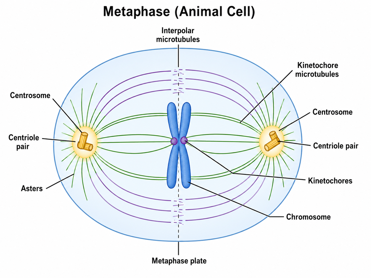
Must knowThe centrosome is the main microtubule-organizing center (MTOC) of animal cells. Each contains a pair of centrioles (cylinders of microtubules) in pericentriolar material. The centrosome duplicates around G1/S so that by G2 there are two; at mitotic onset they migrate to opposite poles and nucleate the spindle. Asters are the star-shaped microtubule arrays radiating from each centrosome, anchoring it to the cell cortex.
Passage-levelPlant cells lack centrioles but still build a functional spindle from other MTOCs — the MCAT may ask what structure is absent in plant mitosis.
The Mitotic Spindle
Must knowThe mitotic spindle is a bipolar microtubule array built from the two centrosomes, with three classes:
- Kinetochore microtubules (K-fibers): attach at kinetochores; move chromosomes
- Polar (interpolar) microtubules: overlap at the equator; push poles apart
- Astral microtubules: reach the cell cortex; anchor the spindle
Chromatids, Centromeres, and Kinetochores
Must knowAfter replication, each chromosome is two identical sister chromatids held together by cohesin. The centromere is the constricted DNA region where the chromatids are joined. On it sits the kinetochore, a protein complex that binds spindle microtubule plus-ends. Proper biorientation (the two kinetochores attached to opposite poles, under tension) is what the spindle assembly checkpoint monitors.
Quick check: What is the difference between a centromere and a kinetochore?
Answer: The centromere is the DNA region; the kinetochore is the protein complex on top of it that binds spindle microtubules. One is DNA, the other is protein.
The Mitotic Phases in Detail
Mitosis is abbreviated PMAT (Prophase, Metaphase, Anaphase, Telophase), but the MCAT also expects prometaphase. Chromosomes condense, are captured by the spindle, align, separate, and then the cell divides.
Prophase
Must know- Chromatin condenses into visible chromosomes (each already two sister chromatids from S phase).
- The spindle begins to form as centrosomes migrate to opposite poles.
- The nucleolus disappears; the nuclear envelope is still intact.
Prometaphase
Must know- Nuclear envelope breaks down (NEBD): MPF phosphorylates lamins, depolymerizing the lamina; the envelope fragments into the ER.
- Spindle microtubules now reach and attach to kinetochores (kinetochore capture).
- Chromosomes are pulled toward the equator.
Metaphase
Must know- All chromosomes align at the metaphase plate (cell's equator), under equal tension from both poles.
- The spindle assembly checkpoint is satisfied only when every kinetochore is under tension; until then the MCC inhibits APC/C.
- Metaphase is used for karyotyping (chromosomes maximally condensed).
Anaphase
Must knowTriggered when APC/C is fully activated:
- APC/C degrades securin → free separase cleaves cohesin.
- Anaphase A: sister chromatids (now individual chromosomes) move to opposite poles via shortening of kinetochore microtubules.
- Anaphase B: the poles move farther apart as polar microtubules slide and elongate.
Each pole ends with a complete 2N set, each chromosome now a single chromatid.
Telophase and Cytokinesis
Must know- Chromosomes arrive at the poles and decondense.
- Nuclear envelopes reassemble (lamins dephosphorylated) and nucleoli reappear; the spindle disassembles.
- Cytokinesis begins in late anaphase/telophase:
- Animal cells: a cleavage furrow from a contractile ring of actin and myosin II pinches the cell in two.
- Plant cells: a cell plate forms from Golgi-derived vesicles, becoming the new cell wall.
Each daughter cell: 2N chromosomes, each a single chromatid.
Quick check: A drug prevents cohesin cleavage. Which mitotic event is blocked?
Answer: The onset of anaphase. Without cohesin cleavage by separase, sister chromatids cannot separate.
Mechanisms of Chromosome Movement
Must knowMicrotubules are dynamic polymers of α/β-tubulin dimers that grow and rapidly depolymerize from their plus-ends. Chromosomes move by two complementary mechanisms:
1. Kinetochore microtubule depolymerization (poleward movement). As K-fibers shorten at the kinetochore, they reel the chromosome toward the pole — the heart of anaphase A.
2. Spindle pole separation. In anaphase B, overlapping polar microtubules slide and elongate so the poles move apart. Know the logic depolymerization pulls chromatids poleward; pole separation drives them apart (motor proteins power this, but names aren't required).
Spindle poisons (high-yield for pharmacology):
- Colchicine / nocodazole: bind tubulin and prevent polymerization → no spindle → M arrest. (Colchicine is used for gout.)
- Taxol (paclitaxel): stabilizes microtubules, preventing depolymerization → spindle can't move chromosomes → M arrest. (Used as chemotherapy.)
Both exploit the spindle assembly checkpoint and lead to mitotic arrest and apoptosis in dividing cancer cells.
Quick check: Both colchicine and taxol arrest cells in mitosis, but by opposite mechanisms. Explain.
Answer: Colchicine prevents polymerization, so no spindle forms. Taxol prevents depolymerization, so the spindle is "frozen" and useless. Both leave kinetochores without proper tension, keeping the spindle assembly checkpoint active.
Nuclear Membrane Breakdown and Reorganization
Must knowThe nuclear lamina is a meshwork of lamin proteins underlying the inner nuclear membrane; it supports the envelope and anchors chromatin and nuclear pores.
Breakdown (prometaphase): MPF phosphorylates lamins → lamina depolymerizes, pores disassemble, and the envelope membrane is absorbed into the ER. This is an irreversible commitment to mitosis.
Reorganization (telophase): As MPF falls (cyclin degraded by APC/C), phosphatases dephosphorylate lamins → the lamina re-polymerizes, ER membranes re-wrap the chromosomes, and pores re-insert.
Why it matters: The MCAT asks what triggers breakdown (MPF) or what happens if lamins can't be phosphorylated (no breakdown → mitosis blocked in prophase/prometaphase).
Quick check: MPF activity is high in M phase and low at the end. What happens to lamin phosphorylation as MPF drops, and what event does this correspond to?
Answer: Lamins are dephosphorylated, allowing them to re-polymerize. This is nuclear envelope reassembly in telophase.
Cancer: Loss of Cell Cycle Controls
Cancer cells accumulate mutations that override checkpoints, allowing unlimited proliferation.
Hallmarks Relevant to Cell Division
Must know- Loss of contact inhibition: normal cells stop dividing when they sense neighbors; cancer cells pile up into disorganized masses.
- Anchorage independence: cancer cells can grow without ECM attachment (tested via soft agar colony assays).
- Replicative immortality: normal cells have a division limit (telomere shortening); cancer cells upregulate telomerase.
- Evasion of apoptosis: loss of p53 lets cells with damaged DNA survive and divide.
- Genomic instability: defective checkpoints let unrepaired DNA accumulate further mutations.
Key Molecular Players in Cancer
Must know| Molecule | Normal Function | Alteration | Mutation Type |
|---|---|---|---|
| Rb | Inhibits E2F; gates G1→S | Inactivated | Loss-of-function, recessive |
| p53 | Senses DNA damage; induces p21 or apoptosis | Lost/mutated | Loss-of-function, recessive |
| Ras | GTPase; transduces growth signals | Stuck "on" | Gain-of-function, dominant |
Oncogenes vs Tumor Suppressors: The Key Distinction
Must knowAn oncogene is a stuck accelerator — constitutively active, so one mutant allele drives cancer (dominant). A tumor suppressor is the brake — both copies must be inactivated (recessive), explaining the two-hit hypothesis for familial cancers like retinoblastoma.
Quick check: A patient inherits one defective RB1 copy. Why does retinoblastoma develop only if the second copy is also mutated in a retinal cell?
Answer: Rb is a recessive tumor suppressor — one functional copy still restrains E2F. Only when the second allele is lost ("second hit") is the brake fully released, allowing uncontrolled proliferation.
Common Confusions & Tricks
1. "Chromosome number" vs. "DNA content" after S phase.
After S phase you still have 2N = 46 chromosomes, but each has two sister chromatids, so DNA content is 4N. Chromosome number stays 46 until anaphase pulls chromatids apart.
2. Centromere ≠ kinetochore.
The centromere is chromosomal DNA; the kinetochore is the protein complex on it. Microtubules attach to the kinetochore.
3. Centrioles ≠ centromere.
Centrioles are cylindrical organelles in the centrosome (MTOC). Centromeres are chromosomal DNA sequences. Easily confused.
4. Colchicine vs. Taxol — opposite mechanisms, same arrest.
Both arrest at M phase. "Spindle absent" → colchicine/nocodazole. "Spindle present but static" → taxol.
5. Mitosis separates sister chromatids; meiosis I separates homologs.
In mitosis, sister chromatids separate in anaphase. In meiosis I, homologous chromosomes separate. Meiosis II looks like mitosis.
6. G0 is not the same as G1.
A G0 cell has actively exited the cycle. Permanent G0 = terminal differentiation (neurons, muscle); reversible G0 = quiescence. Cells don't "sit in G1 indefinitely."
7. Anaphase A vs. Anaphase B.
A = chromosomes move to poles (K-fibers shorten). B = poles move apart (polar microtubules elongate/slide).
8. Rb is inactive when phosphorylated.
Counterintuitive: Rb is active (binds E2F) when hypophosphorylated; phosphorylation by a G1 cyclin–CDK inactivates it (releases E2F → S phase).
9. Plant cells lack centrioles but still complete mitosis.
Plants have no centrioles or asters but still form a bipolar spindle and use a cell plate (not a cleavage furrow).
10. The spindle assembly checkpoint monitors TENSION, not just attachment.
Attachments that don't generate tension still activate the checkpoint. Only proper biorientation under tension satisfies it.
Key Takeaways
Cell Cycle Phases
- G1: Growth; restriction point; Rb/E2F central
- S: DNA replication; DNA doubles 2N→4N; chromosome number stays 2N
- G2: Final prep; G2/M checkpoint
- M: Mitosis (PMAT + prometaphase) + cytokinesis; each daughter back to 2N
- G0: Quiescent (reversible) or terminally differentiated (irreversible) exit
Mitotic Stages
- Prophase: chromatin condenses; spindle forms; nucleolus disappears
- Prometaphase: nuclear envelope breaks down (MPF phosphorylates lamins); kinetochore capture
- Metaphase: chromosomes aligned under tension; spindle checkpoint satisfied
- Anaphase: cohesin cleaved by separase → chromatids separate (A: to poles; B: poles apart)
- Telophase: decondensation; envelope reassembly; nucleolus returns
- Cytokinesis: cleavage furrow (animals) or cell plate (plants)
Key Structures
- Centrosome (centriole pair): MTOC; nucleates spindle; duplicates in S
- Kinetochore: protein complex on centromere; microtubule attachment site
- Cohesin: holds sister chromatids; cleaved by separase at anaphase
- Asters: anchor centrosomes; establish division axis
Cell Cycle Regulators
- CDKs + Cyclins: drive progression; key complex = MPF for mitotic entry
- Rb: tumor suppressor; inhibits E2F when hypophosphorylated
- p53: "guardian of the genome"; induces p21 or apoptosis on DNA damage
- APC/C: activated at metaphase→anaphase; destroys mitotic cyclin and securin
Checkpoints
- G1/S (restriction point): DNA damage? p53 → p21 → CDK inhibition
- G2/M: replication complete? DNA repaired?
- Spindle Assembly Checkpoint: all kinetochores bioriented under tension? MCC inhibits APC/C until satisfied
Cancer
- Oncogenes: gain-of-function; dominant; stuck "on"
- Tumor suppressors: loss-of-function; recessive; two-hit model (Rb, p53)
- Loss of: contact inhibition, anchorage dependence, apoptosis, replicative limit (telomerase up)
Spindle Poisons (High-Yield)
- Colchicine/nocodazole: prevent polymerization → no spindle → M arrest
- Taxol: prevents depolymerization → frozen spindle → M arrest