The reproductive system tells a sequential story from gamete creation through birth. Understanding the logic of each step (why does meiosis arrest here? why does the egg keep more cytoplasm?) lets you reason through novel passages rather than memorize.
Priority labels: Must know = cold; Know the logic = mechanism not names; Passage-level = recognize, don't memorize; Optional = skippable.
Gametogenesis by Meiosis
The Purpose and Logic of Meiosis
Must knowSexually reproducing organisms must halve their chromosome number before combining two gametes, or each generation would double the count. Meiosis does this in two divisions: meiosis I (reductional — separates homologous chromosomes) and meiosis II (equational — separates sister chromatids, like mitosis).
A cell entering meiosis is 2n (diploid) with replicated DNA. After meiosis I, each daughter is 1n (haploid) but chromatids are still paired. After meiosis II, each cell is fully haploid with single chromatids — these are the gametes.
Three sources of genetic variation:
- Crossing over (recombination) in prophase I, when homologs synapse into bivalents/tetrads (chiasmata mark crossover points).
- Independent assortment of homologs at metaphase I ( combinations in humans).
- Random fertilization — any sperm can fertilize any egg, multiplying the diversity.
Quick check: A cell entering meiosis has 46 chromosomes (23 pairs). How many chromosomes are in each cell immediately after meiosis I completes? How many after meiosis II?
Answer: After meiosis I: 23 chromosomes, each consisting of two sister chromatids (the cell is haploid but chromatids are still joined). After meiosis II: 23 chromosomes, each a single chromatid — fully haploid gametes.
Spermatogenesis
From Stem Cell to Spermatozoon
Must knowSpermatogenesis is a continuous process occurring from puberty throughout life, taking place in the seminiferous tubules of the testes. The pathway is:
Spermatogonia (stem cells) divide mitotically: some self-renew, others become primary spermatocytes that enter meiosis. Meiosis I → two secondary spermatocytes; meiosis II → four spermatids; spermiogenesis (morphological transformation, not a division) → mature spermatozoa.
Critically, one primary spermatocyte → four functional sperm. This 1:4 ratio is a key contrast with oogenesis.
Supporting Cells of the Testes
Must know- Sertoli cells (inside tubules): nurse developing sperm and form the blood-testis barrier that protects the haploid cells.
- Leydig (interstitial) cells (outside tubules): produce testosterone, which drives spermatogenesis.
(The hormonal regulation of these cells — FSH/LH, inhibin, and the male HPG axis — is detailed in the 3B Reproductive System guide.)
Morphology of the Mature Sperm
Must knowThe spermatozoon's structure reflects its single mission — delivering paternal DNA to the egg:
| Region | Structure | Function |
|---|---|---|
| Head | Nucleus (condensed DNA) + acrosome cap | Carries haploid genome; acrosome stores hydrolytic enzymes (hyaluronidase, proteases) for egg penetration |
| Midpiece | Densely packed mitochondria around axoneme | ATP production via oxidative phosphorylation for flagellar motility |
| Tail (flagellum) | 9+2 axoneme (microtubule arrangement) | Propulsion |
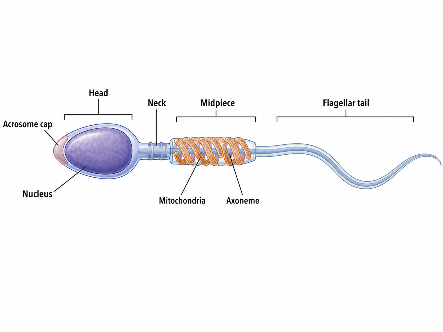
Mature sperm shed almost all cytoplasm during spermiogenesis, so the paternal (midpiece) mitochondria are typically degraded after fertilization (see Relative Contribution).
Quick check: A student claims that sperm motility depends primarily on glycolysis because the sperm head contains glycolytic enzymes. What's wrong with this claim?
Answer: Sperm motility is powered primarily by ATP from oxidative phosphorylation in the midpiece mitochondria, not glycolysis in the head.
Oogenesis
A Story of Strategic Arrest
Must knowOogenesis is fundamentally different from spermatogenesis in timing, yield, and continuity. The entire process is characterized by two strategic arrests that the MCAT tests repeatedly.
Arrest 1 — Prophase I: All primary oocytes form by fetal life. A female is born with her entire lifetime supply (~1–2 million), all arrested in prophase I, until puberty. Each month FSH recruits a cohort and one dominant follicle progresses.
Just before ovulation, the LH surge triggers completion of meiosis I → one secondary oocyte (1n, keeps most cytoplasm) + one first polar body (small, often degenerates).
Arrest 2 — Metaphase II: The secondary oocyte enters meiosis II but arrests in metaphase II. This is the cell that is ovulated; it completes meiosis II only if a sperm fertilizes it, producing the ovum + a second polar body.
Tally: one primary oocyte → one functional ovum + up to three polar bodies (all degenerate). Versus spermatogenesis: one primary spermatocyte → four sperm.
Follicular Development
Know the logicThe oocyte matures inside follicular structures; track the progression, don't memorize every cell layer:
- Primordial → primary → secondary → Graafian follicle: granulosa cells proliferate, the zona pellucida (glycoprotein coat) forms, theca cells differentiate, and a fluid-filled antrum enlarges; the dominant Graafian follicle completes meiosis I just before ovulation.
- Corpus luteum: the post-ovulation follicular remnant. If implantation occurs, hCG (from the trophoblast) sustains it; otherwise it regresses to the corpus albicans. Its endometrial/hormonal role in the cycle is covered in the 3B Reproductive System guide.
Hormonal Triggers (in brief)
Must knowFor gametogenesis, the key cue is that the LH surge triggers ovulation and completion of meiosis I; the secondary oocyte is then released. The full HPG axis, the FSH/LH feedback loops, and the follicular/luteal phases of the menstrual cycle are covered in the 3B Reproductive System guide.
Quick check: A woman has a mutation causing her secondary oocytes to fail to arrest in metaphase II and instead complete meiosis II immediately after meiosis I. What consequence would you expect?
Answer: The oocyte would finish meiosis II before fertilization, becoming a "finished" ovum with no sperm-triggered completion step. The metaphase II arrest is a checkpoint that ties completion of meiosis to sperm entry; losing it tends to yield a less developmentally competent egg.
Differences in Formation: Spermatogenesis vs. Oogenesis
Must knowOne of the highest-yield comparisons on the MCAT:
| Feature | Spermatogenesis | Oogenesis |
|---|---|---|
| Onset | Puberty | Fetal life |
| Continuity | Continuous, lifelong | Discontinuous; arrested twice |
| Arrest point(s) | None | Prophase I (until ovulation); Metaphase II (until fertilization) |
| Yield from one primary cell | 4 functional sperm | 1 functional egg + up to 3 polar bodies |
| Location | Seminiferous tubules | Ovarian follicles |
| Completion trigger | Autonomous | Meiosis I by LH surge; meiosis II by fertilization |
The asymmetric yield is logical: the egg must be large and packed with maternal cytoplasm (ribosomes, mRNA, organelles), so oogenesis concentrates resources into one cell by offloading chromosomes (with minimal cytoplasm) into polar bodies. Sperm are tiny and disposable — their only job is to carry DNA.
Quick check: Why are polar bodies produced rather than simply discarding chromosomes by another mechanism?
Answer: Meiosis inherently requires nuclear division. The polar bodies are the inevitable byproduct of completing chromosomal reduction while retaining maximal cytoplasm in one cell. Each "discard" cell still receives a haploid nucleus because meiosis doesn't have a mechanism to selectively distribute chromosomes without cell division.
Differences in Morphology
Sperm vs. Egg: Form Follows Function
Must knowThe sperm is essentially a motile delivery capsule for haploid DNA:
- Highly condensed chromatin in the head (histones replaced by protamines)
- Acrosome — lysosome-like cap; hydrolytic enzymes for penetrating egg coats
- Midpiece — mitochondria for ATP
- Flagellum with the classic 9+2 axoneme, powered by dynein
The egg (secondary oocyte at ovulation) is one of the largest human cells:
- Zona pellucida: glycoprotein coat; ZP3 is the sperm receptor that triggers the acrosomal reaction
- Cortical granules: beneath the membrane; released after fertilization to harden the zona (slow block to polyspermy)
- Corona radiata: layer of granulosa cells around the zona; sperm penetrate it with hyaluronidase
- Large cytoplasm with maternal ribosomes, mRNA, organelles, and stores
- No flagellum — non-motile; moved by fallopian tube cilia and contractions
Quick check: What would happen to fertilization if a sperm had a genetic defect causing absent acrosomal enzymes?
Answer: The sperm would be able to bind the zona pellucida (binding depends on surface glycoproteins interacting with ZP3) but would fail to penetrate it. Without the hydrolytic enzymes from the acrosome (hyaluronidase, acrosin/proteases), the sperm cannot digest a path through the zona pellucida and cumulus cells. Fertilization would not occur despite normal binding.
Relative Contribution to the Next Generation
What Each Gamete Actually Contributes
Must knowThe MCAT probes what is inherited from each parent:
Paternal (sperm):
- Haploid nuclear DNA (22 autosomes + X or Y; Y-bearing sperm determines male sex)
- A centriole the zygote uses to organize its first mitotic spindle (the oocyte supplies no functional centriole)
- Essentially no cytoplasm or persistent organelles
Maternal (egg):
- Haploid nuclear DNA (22 autosomes + X)
- Virtually all cytoplasm
- All functional mitochondria and mtDNA → basis of maternal (mitochondrial) inheritance; paternal mitochondria are degraded after fertilization
- Maternal mRNAs/proteins (made during oogenesis) that direct early development before the embryonic genome activates
- Ribosomes and cytoplasmic machinery
So early embryonic development is run largely by maternal gene products until the embryonic genome takes over.
Quick check: A woman has a mitochondrial mutation causing a mitochondrial disease. She and her husband have three children. Which children, if any, would you expect to inherit the disease?
Answer: All three children would be at risk of inheriting the disease, regardless of sex, because mitochondria are inherited maternally. The father's mitochondria are degraded after fertilization. (Note: penetrance and expressivity of mitochondrial diseases can vary due to heteroplasmy — not all mitochondria in a cell need to carry the mutation — but the inheritance pattern is strictly maternal.)
Reproductive Sequence: Fertilization, Implantation, Development, and Birth
Fertilization
Must knowFertilization normally occurs in the ampulla of the fallopian tube. Sperm travel from the vagina through the cervix and uterus, undergoing capacitation on the way.
Capacitation: sperm maturation in the female tract (~hours) — membrane changes that enable hyperactivated motility and acrosome-reaction competence. Sperm cannot fertilize immediately upon ejaculation.
Steps of fertilization (protein names are lower-yield — recognize, don't memorize):
- Sperm penetrates corona radiata/cumulus using acrosomal hyaluronidase.
- Sperm binds zona pellucida (ZP3), triggering the acrosomal reaction: the acrosome releases proteases (acrosin) that digest a path through the zona.
- Sperm-egg membrane fusion (mediated by sperm IZUMO1 + egg JUNO).
- Blocks to polyspermy (polyspermy → nonviable triploid zygote):
- Fast block: within seconds, the egg membrane depolarizes (~−70 → +20 mV), transiently blocking additional sperm. Electrical, temporary.
- Slow block (cortical/zona reaction): within minutes, cortical granules exocytose enzymes that harden the zona pellucida. Chemical, permanent.
- Completion of meiosis II: sperm entry triggers the secondary oocyte to finish meiosis II (→ ovum + second polar body). The female and male pronuclei do not fuse — they break down their envelopes and their chromosomes congress on a shared spindle for the first mitotic division.
Quick check: A researcher develops a drug that blocks cortical granule exocytosis in mouse eggs. What is the predicted outcome when these eggs are fertilized in vitro?
Answer: The fast block (membrane depolarization) would still occur temporarily, but without the slow block (cortical reaction), the zona pellucida would not be hardened. Multiple sperm could penetrate the zona and fuse with the egg, causing polyspermy — a triploid or aneuploid zygote that typically fails to develop normally.
Cleavage and Early Development
Must knowAfter fertilization, the zygote undergoes cleavage — rapid mitotic divisions with no growth between them, so total volume stays constant while blastomeres get progressively smaller.
Sequence: zygote → morula (solid ball, ~day 3–4) → blastocyst (hollow, ~day 4–5) → implantation (days 6–10) → gastrulation (~weeks 2–3) → neurulation (~weeks 3–4).
Blastocyst structure:
- Trophoblast: outer layer → placenta and extraembryonic membranes; secretes hCG, which maintains the corpus luteum (and thus progesterone) in early pregnancy. hCG is what pregnancy tests detect.
- Inner cell mass (ICM/embryoblast): → the embryo proper (plus amnion/yolk sac).
- Blastocoel: fluid-filled cavity.
Quick check: Why would a drug that blocks hCG secretion immediately after implantation cause pregnancy loss?
Answer: Without hCG, the corpus luteum receives no maintenance signal and degenerates (as it would in a normal non-pregnant cycle). The corpus luteum is the primary source of progesterone in early pregnancy. Progesterone maintains the uterine endometrium in a secretory, receptive state. Loss of progesterone would cause endometrial shedding, carrying the implanted embryo with it — i.e., early pregnancy loss. (This is the mechanism exploited by some hormonal contraceptive strategies.)
Implantation
Must knowThe blastocyst hatches from the zona (~day 4–5) and adheres to the uterine endometrium (kept secretory by progesterone from the corpus luteum); implantation occurs ~days 6–10.
Passage-levelThe trophoblast splits into an inner cytotrophoblast and an outer invasive syncytiotrophoblast (secretes hCG) that erodes into maternal vessels, establishing the maternal-fetal interface.
Gastrulation, Germ Layers, and Neurulation (briefly)
Must knowGastrulation establishes the three germ layers (ectoderm, mesoderm, endoderm) via migration through the primitive streak; neurulation follows, in which the notochord induces the overlying ectoderm to form the neural tube (future CNS). Germ-layer derivatives and neural tube defects are covered in the Embryogenesis / Mechanisms of Development guides.
The Placenta
Must knowThe placenta exchanges gases, nutrients, and wastes between fetal and maternal blood without the two mixing. It is also endocrine: it secretes estrogen, progesterone (taking over from the corpus luteum partway through pregnancy — the luteal-placental shift), and hCG.
Birth (Parturition)
Must knowParturition concludes development by expelling the fetus and placenta.
- Oxytocin drives uterine contractions via a positive feedback loop: contractions → cervical stretch → more oxytocin → stronger contractions.
- Three stages of labor: (1) cervical dilation, (2) fetal expulsion, (3) placental delivery.
(The full hormonal control of parturition — the estrogen:progesterone ratio shift, prostaglandins, and relaxin — is covered in the 3B Reproductive System guide.)
Quick check: A patient is given a drug that blocks oxytocin receptors in the uterus. What would you predict happens to uterine contractions during labor?
Answer: Uterine contractions would be significantly diminished or halted, because oxytocin is the primary driver of myometrial contractions during labor. The positive feedback loop would be broken — cervical pressure would fail to amplify contractions. This is clinically relevant: oxytocin receptor antagonists (like atosiban) are used as tocolytics to stop preterm labor.
Common Confusions & Tricks
1. "Arrested in what phase?" is a classic MCAT trap.
Primary oocytes arrest in prophase I (not metaphase I, not prophase II). Secondary oocytes arrest in metaphase II (not metaphase I). If a question describes an oocyte that has just been ovulated, it is a secondary oocyte in metaphase II — not yet a mature ovum. The word "ovum" technically refers to the cell after fertilization triggers completion of meiosis II. Many students lose points by thinking ovulation releases a "mature egg."
2. Spermatogenesis has no arrest; oogenesis has two.
Students sometimes apply the concept of meiotic arrest to sperm — spermatocytes proceed through meiosis I and II without pause. Arrests are an exclusive feature of oogenesis.
3. The 1:4 vs. 1:1 ratio trap.
One primary spermatocyte → 4 functional sperm. One primary oocyte → 1 functional egg + polar bodies. Don't confuse polar bodies with functional gametes.
4. Trophoblast vs. inner cell mass.
The trophoblast → placenta and hCG (extraembryonic). The inner cell mass → embryo and amnion/yolk sac. Students frequently reverse these. Remember: tropho- means nourishment/feeding → the trophoblast feeds the embryo by forming the placenta.
5. hCG maintains the corpus luteum; it does NOT replace progesterone directly.
hCG acts like LH to keep the corpus luteum alive. It is the corpus luteum that then secretes progesterone. Pregnancy tests detect hCG, not progesterone.
6. Maternal mitochondria, always.
Paternal mitochondria are tagged for destruction after fertilization. All of your mitochondria (and mtDNA) came from your mother. If the MCAT describes a mitochondrial disease, the inheritance is strictly maternal — it passes to all offspring (sons and daughters), but sons do not pass it on to their children.
7. Fast block is electrical and temporary; slow block is chemical and permanent.
If a question asks about the immediate block to polyspermy, it is the fast block (membrane depolarization). If it asks about the lasting structural change, it is the slow block (cortical/zona reaction). Both must occur for normal fertilization.
8. The fallopian tube is where fertilization happens, not the uterus.
Fertilization occurs in the ampulla of the fallopian tube. The zygote then travels toward the uterus over ~5–7 days while developing into a blastocyst before implanting. If implantation occurs in the tube, that is an ectopic pregnancy — a surgical emergency.
Key Takeaways
Gametogenesis Comparison
| Spermatogenesis | Oogenesis | |
|---|---|---|
| Timing of onset | Puberty | Fetal life |
| Meiotic arrest | None | Prophase I → Metaphase II |
| Yield | 4 sperm per primary spermatocyte | 1 egg (+ polar bodies) per primary oocyte |
| Continuity | Lifelong, continuous | Cyclic; all oocytes formed before birth |
| Supporting cells | Sertoli (FSH) + Leydig (LH → testosterone) | Granulosa (FSH) + theca cells (LH → androgens) |
Sperm Morphology
- Head: condensed nucleus + acrosome (lysosome-like, hydrolytic enzymes)
- Midpiece: mitochondria (energy for motility)
- Tail: 9+2 axoneme, dynein-powered
Egg Morphology
- Zona pellucida (ZP3 = sperm receptor); cortical granules (slow block); large cytoplasm with maternal stores
Maternal vs. Paternal Contribution
- Sperm: haploid nuclear DNA (plus a centriole for the first spindle)
- Egg: haploid nuclear DNA + ALL cytoplasm + ALL functional mitochondria (mtDNA is maternally inherited)
Fertilization Sequence
- Capacitation in female tract
- Sperm penetrates cumulus (hyaluronidase)
- Sperm binds ZP3 → acrosomal reaction
- Sperm-egg membrane fusion → fast block (depolarization, seconds) → slow block (cortical reaction, minutes)
- Secondary oocyte completes meiosis II → female pronucleus forms
- Pronuclei congress on mitotic spindle → first cleavage
Early Development Sequence
- Zygote → cleavage → morula → blastocyst (trophoblast + ICM + blastocoel) → implantation (days 6–10)
- Trophoblast → placenta + hCG; ICM → embryo proper
- Gastrulation (three germ layers) and neurulation follow — detailed in the Embryogenesis / Mechanisms of Development guides
- Placenta: exchanges gases/nutrients/wastes (fetal and maternal blood do not mix); secretes estrogen, progesterone, hCG
Birth
- Positive feedback loop: oxytocin ↔ uterine contractions
- Three stages: cervical dilation → fetal expulsion → placental delivery
- Hormonal control of labor: see the 3B Reproductive System guide