Introduction: Why Neurons Are Worth Understanding Deeply
Neurons are the signaling units of the nervous system. Everything from a reflex to conscious thought relies on the same core mechanisms: maintaining an electrochemical gradient, firing an electrical signal, and chemically communicating across a gap. The MCAT tests your ability to reason about what happens when a channel opens, a pump is blocked, or excitatory and inhibitory inputs arrive together. Build the intuition here and these questions become straightforward.
Priority labels: Must know = cold; Know the logic = mechanism not names; Passage-level = recognize, don't memorize; Optional = skippable.
Neuron Anatomy: The Cell Body and Its Extensions
The Cell Body (Soma)
Must knowThe soma (cell body) is the neuron's metabolic headquarters: it holds the nucleus and the organelles for protein synthesis and energy production (key for the ATP-demanding ion pumping).
Most adult neurons are post-mitotic (do not divide), which is why neural damage from stroke, neurodegeneration, or trauma tends to be permanent.
The soma integrates incoming signals from dendrites and, if the combined input reaches threshold, initiates an action potential at the axon hillock (where the axon leaves the soma).
Quick check: Why is the axon hillock — not the middle of the axon — the site where action potentials are initiated?
Answer: The axon hillock has the highest density of voltage-gated sodium channels of any part of the neuron. It is the most excitable location, so it reaches threshold first when depolarizing current spreads from the dendrites and soma inward.
Dendrites
Must knowDendrites are branched extensions of the soma that receive incoming signals. Their branching increases surface area for synaptic input. They conduct signals toward the cell body, carrying graded potentials — local changes in voltage whose magnitude reflects stimulus strength. Graded potentials spread passively and decay with distance, which is why integration at the axon hillock (where these decaying signals sum) is the decision point.
OptionalDendritic spines are sites of excitatory synapses; changes in their number/shape underlie synaptic plasticity and memory.
Quick check: A neurotransmitter binding to a dendritic receptor causes a small depolarization. Will this depolarization be larger or smaller by the time it reaches the axon hillock, compared to at the synapse itself?
Answer: Smaller. Graded potentials decay with distance because they spread passively through cytoplasm, which has resistance. This is why a single small input rarely triggers an action potential — multiple inputs must sum.
The Axon: Structure and Function
Must knowThe axon is the output cable of the neuron. Key features:
- Axon hillock: origin of the axon; site of action potential initiation.
- Axon: conducts action potentials away from the soma; a neuron has one axon that may branch into collaterals.
- Axon terminal (synaptic bouton): the swollen ending containing synaptic vesicles of neurotransmitter.
Conduction velocity depends on two factors: axon diameter (larger = less resistance = faster) and myelination (next section).
Quick check: In which direction does the action potential normally travel along an axon?
Answer: From the axon hillock toward the axon terminal (anterograde, i.e., away from the soma). This directionality is enforced by the refractory period — the region just behind the advancing impulse is briefly inexcitable, preventing backward propagation.
Myelin Sheath, Schwann Cells, and Axon Insulation
The Myelin Sheath
Must knowThe myelin sheath is a lipid-rich, multilayered wrapping (like tape on a wire) that insulates the axon and dramatically increases conduction velocity. It forces ionic current to flow along the axon interior rather than leaking out across the membrane between nodes, making propagation far more efficient.
Schwann Cells vs. Oligodendrocytes
Must know| Feature | Schwann Cells | Oligodendrocytes |
|---|---|---|
| Location | PNS | CNS |
| Myelinate | One axon segment per cell | Multiple segments per cell |
| Regeneration | Support axon regeneration | Do NOT support regeneration |
This is why PNS injuries (severed peripheral nerve) recover better than CNS injuries (spinal cord): Schwann cells provide a scaffold and growth factors that guide regenerating axons; oligodendrocytes cannot.
Passage-levelClinical connection: Multiple sclerosis is an autoimmune attack on CNS myelin; demyelination slows or blocks conduction, causing relapsing neurological symptoms.
Quick check: A patient with MS has a lesion demyelinating a visual pathway. Would you expect conduction velocity in that pathway to increase, decrease, or be unaffected?
Answer: Decrease (slow down). Myelin is essential for rapid saltatory conduction. Without myelin, the axon either conducts slowly via continuous conduction or fails to conduct at all if the damage is severe.
Nodes of Ranvier and Saltatory Conduction
Must knowNodes of Ranvier are gaps between myelin segments where the axon membrane is exposed and densely packed with voltage-gated Na⁺ channels (these channels are absent under the myelinated internodes).
Saltatory conduction: rather than depolarizing every patch of membrane in sequence (slow, continuous conduction in unmyelinated axons), the action potential "jumps" from node to node. Current from a firing node flows down the low-resistance axon interior to the next node, where it triggers a fresh action potential. This is both faster and more energy-efficient (the Na⁺/K⁺ ATPase only needs to restore gradients at the nodes, not along the whole axon).
Quick check: If a toxin specifically blocked voltage-gated Na⁺ channels at one node of Ranvier, what would happen to action potential propagation?
Answer: Propagation would fail at that node. The action potential cannot "jump over" a blocked node because the arriving current would be insufficient to depolarize the next node beyond threshold. The signal would be extinguished — this is analogous to the conduction block seen in local anesthesia (e.g., lidocaine blocks voltage-gated Na⁺ channels).
The Synapse: Site of Impulse Propagation Between Cells
Synapse Architecture
Must knowA synapse is the junction between a neuron and its target (another neuron or an effector like muscle). Most are chemical synapses, which convert the electrical signal to a chemical message. Three components:
- Presynaptic terminal: contains synaptic vesicles of neurotransmitter.
- Synaptic cleft: the narrow extracellular gap between cells.
- Postsynaptic membrane: bears receptors that bind the neurotransmitter.
Electrical synapses (gap junctions) allow direct ionic current flow between cells — faster and bidirectional, but rare in the mammalian CNS (common in cardiac/smooth muscle).
How the Chemical Synapse Transmits a Signal
Must know- An action potential reaches the axon terminal.
- Voltage-gated Ca²⁺ channels open.
- Ca²⁺ influx triggers exocytosis of vesicles → neurotransmitter released into the cleft.
- Neurotransmitter diffuses across and binds postsynaptic receptors.
- Neurotransmitter is cleared by reuptake, enzymatic degradation, or diffusion.
Signaling is one-way (presynaptic → postsynaptic) because only the presynaptic side releases transmitter and only the postsynaptic side has receptors.
Quick check: A drug blocks voltage-gated Ca²⁺ channels at axon terminals. What effect would you predict on neurotransmitter release?
Answer: Neurotransmitter release would be drastically reduced or abolished. Ca²⁺ influx is the direct trigger for vesicle fusion and exocytosis. Without Ca²⁺, the action potential arrives but no transmitter is released.
Synaptic Activity: Transmitter Molecules
Must knowNeurotransmitters are the chemical messengers released at synapses. Know these examples and the high-yield distinctions:
| Neurotransmitter | Key Roles / MCAT Relevance |
|---|---|
| Acetylcholine (ACh) | Neuromuscular junction; parasympathetic; degraded by acetylcholinesterase. |
| Dopamine | Reward, motor control; low in Parkinson's. |
| Norepinephrine / Epinephrine | Sympathetic activity; arousal. |
| Serotonin (5-HT) | Mood, sleep, appetite; target of SSRIs. |
| GABA | Major inhibitory CNS transmitter (opens Cl⁻ channels; hyperpolarizes). |
| Glutamate | Major excitatory CNS transmitter. |
| Glycine | Inhibitory in spinal cord. |
Two receptor types:
- Ionotropic (ligand-gated ion channels): fast; channel opens directly on binding (e.g., nicotinic ACh, GABA-A).
- Metabotropic (GPCRs): slow; act via second-messenger cascades (e.g., muscarinic ACh, dopamine receptors).
Quick check: Benzodiazepines (e.g., diazepam) enhance GABA activity at GABA-A receptors. Would you expect the postsynaptic cell to be more or less likely to fire an action potential?
Answer: Less likely. GABA-A is an ionotropic receptor that, when activated, opens Cl⁻ channels. Cl⁻ flows into the cell (down its electrochemical gradient), hyperpolarizing the membrane and making it harder to reach threshold. Benzodiazepines enhance this effect — which is why they are sedating and anti-anxiety.
Resting Potential: The Electrochemical Gradient
Must knowThe resting membrane potential of a typical neuron is about −70 mV (inside negative). It arises from two things working together:
- Concentration gradients (built by the Na⁺/K⁺ ATPase): high K⁺ inside, high Na⁺ (and Cl⁻) outside, plus trapped negative proteins inside.
- Selective permeability: at rest the membrane is highly permeable to K⁺ (leak channels) but not to Na⁺. K⁺ flows out down its gradient, leaving the inside negative; the growing negative charge eventually opposes further K⁺ efflux.
Equilibrium potentials: each ion's equilibrium potential is the voltage at which its electrical pull exactly balances its concentration gradient ( mV, mV). The resting potential (−70 mV) is a permeability-weighted average — close to because the membrane is mostly K⁺-permeable, pulled slightly positive by small Na⁺ permeability. You do not need to compute these here; the Nernst equation is covered in the Electrochemistry guide.
Quick check: If the extracellular K⁺ concentration is suddenly increased (e.g., in hyperkalemia), what happens to the resting membrane potential?
Answer: The cell depolarizes (becomes less negative). With more K⁺ outside, the concentration gradient driving K⁺ out is reduced, so K⁺ equilibrium potential shifts toward 0 mV. The resting potential moves with it. In severe hyperkalemia, neurons and cardiac cells depolarize enough to become dangerously excitable (or paradoxically inexcitable due to inactivation of Na⁺ channels) — this is why hyperkalemia causes cardiac arrhythmias.
The Sodium-Potassium Pump
Must knowThe Na⁺/K⁺ ATPase maintains the concentration gradients underlying the resting potential. Per ATP hydrolyzed, it moves 3 Na⁺ OUT and 2 K⁺ IN. Because it exports more positive charge than it imports, it is electrogenic (a small direct contribution to the negative resting potential), but most of the resting potential comes from K⁺ leak.
Its main role is homeostatic: restoring gradients after repeated action potentials, which would otherwise gradually dissipate.
Passage-levelHigh-yield pharmacology: Cardiac glycosides (digoxin, ouabain) inhibit the Na⁺/K⁺ ATPase; rising intracellular Na⁺ reduces Na⁺/Ca²⁺ exchange, raising intracellular Ca²⁺ and strengthening cardiac contraction.
Quick check: If the Na⁺/K⁺ ATPase is inhibited, what ultimately happens to the resting potential over time?
Answer: The resting potential gradually depolarizes (becomes less negative). Without the pump, Na⁺ leaking in is not removed, so intracellular Na⁺ rises. The Na⁺ concentration gradient dissipates, reducing the driving force for Na⁺ and also altering the K⁺ gradient, ultimately collapsing the resting potential toward 0 mV.
Action Potential
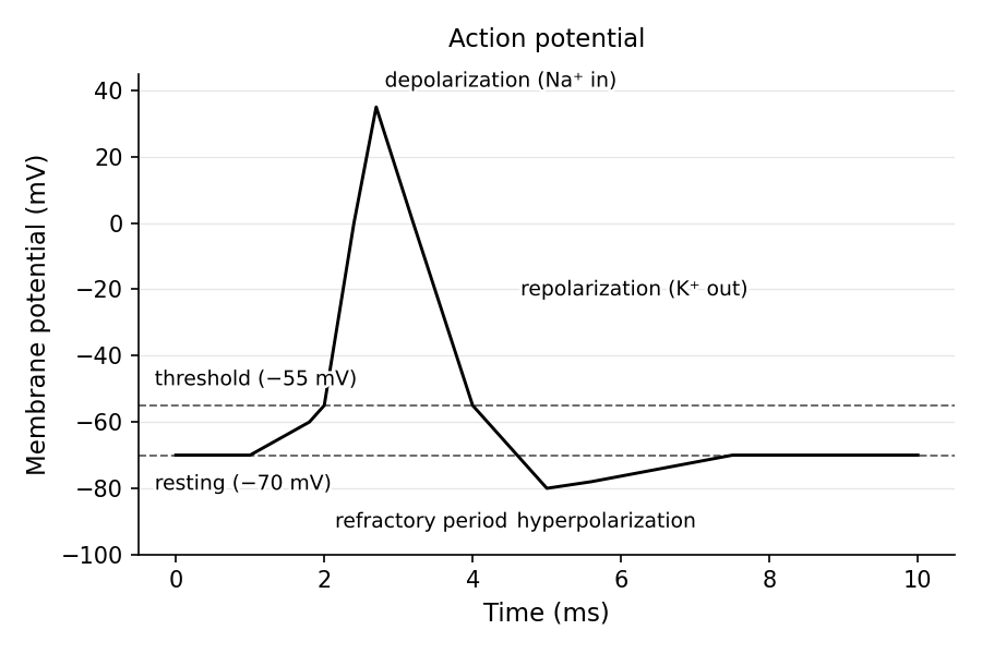
Must knowAn action potential is a rapid, self-propagating reversal and restoration of membrane voltage, driven by voltage-gated Na⁺ channels and voltage-gated K⁺ channels.
Phases of the Action Potential
Must knowKnow the sequence:
- Resting (−70 mV): voltage-gated Na⁺ channels closed; K⁺ leak channels open.
- Depolarization: stimulus reaches threshold (~−55 mV) → voltage-gated Na⁺ channels open → Na⁺ rushes in → membrane swings positive, overshooting to ~+30 mV.
- Repolarization: Na⁺ channels inactivate (cannot reopen until the membrane repolarizes) and voltage-gated K⁺ channels open (more slowly) → K⁺ flows out → membrane drives back negative.
- Hyperpolarization (undershoot): slow K⁺ channel closure lets K⁺ keep leaving, dipping below −70 mV (afterhyperpolarization), then the Na⁺/K⁺ ATPase restores gradients and the membrane returns to rest.
Refractory Periods
Must know- Absolute refractory period: Na⁺ channels are inactivated; no stimulus can fire another action potential. (Corresponds to depolarization/early repolarization.)
- Relative refractory period: Na⁺ channels have recovered but the membrane is hyperpolarized; only a stronger-than-normal stimulus can fire.
Quick check: Why can an action potential not travel backward along a normal axon?
Answer: The absolute refractory period. The region of membrane just behind the advancing action potential has Na⁺ channels in the inactivated state — they cannot reopen in response to the depolarization arriving from the leading edge. Therefore, the impulse can only propagate forward into rested membrane.
Threshold and the All-or-None Principle
Must knowThreshold (~−55 mV) is the voltage at which Na⁺ channel opening becomes self-sustaining: below it, a few channels open but the influx fizzles out; at it, Na⁺ influx opens more channels in a runaway (regenerative) depolarization.
The all-or-none law: once threshold is reached, the action potential fires with the same amplitude and duration regardless of stimulus strength. A stronger stimulus does not produce a bigger action potential. Stimulus intensity is instead encoded by frequency coding (more action potentials per second) and recruitment of more neurons.
Quick check: You pinch someone gently versus hard. Both stimuli trigger action potentials in sensory neurons. How does the nervous system distinguish these two intensities?
Answer: The harder pinch causes (1) a higher frequency of action potential firing in individual neurons and (2) recruitment of additional neurons that have higher thresholds. Both signals (rate and population) convey greater intensity to the CNS.
Excitatory and Inhibitory Nerve Fibers: Summation and Frequency of Firing
EPSPs and IPSPs
Must knowNeurotransmitter binding produces either:
- EPSP: a small depolarization (toward threshold) — e.g., glutamate opening Na⁺/Ca²⁺ channels. Makes firing more likely.
- IPSP: a hyperpolarization (away from threshold) — e.g., GABA opening Cl⁻ channels (Cl⁻ in) or K⁺ channels (K⁺ out). Makes firing less likely.
A single EPSP almost never reaches threshold; the neuron must integrate many inputs.
Spatial and Temporal Summation
Must know- Spatial summation: multiple inputs at different locations arrive simultaneously and sum at the axon hillock.
- Temporal summation: a single input fires repeatedly in rapid succession, so EPSPs add before each fully decays.
The neuron acts as an integrator, summing all EPSPs and IPSPs at the axon hillock to decide whether threshold is reached.
Quick check: A neuron receives 5 EPSPs and 3 IPSPs simultaneously, each of 2 mV magnitude (EPSPs depolarizing, IPSPs hyperpolarizing). The resting potential is −70 mV and threshold is −55 mV. Does the neuron fire?
Answer: Net change = (5 × 2 mV depolarizing) − (3 × 2 mV hyperpolarizing) = +10 mV − 6 mV = +4 mV net depolarization. New membrane potential = −70 + 4 = −66 mV. This does not reach threshold (−55 mV), so no action potential fires. This illustrates how inhibitory inputs can prevent firing even when excitatory inputs are present.
Glial Cells (Neuroglia)
Must knowGlial cells are the non-neuronal support cells of the nervous system; unlike most neurons, they retain the ability to divide. Know the major types and functions:
CNS:
- Astrocytes (most abundant): structural support, form the blood-brain barrier, buffer extracellular K⁺, recycle neurotransmitters (e.g., glutamate).
- Oligodendrocytes: myelinate CNS axons (multiple segments per cell); destroyed in MS.
- Microglia: resident immune cells — the CNS macrophages — phagocytose debris. (Note: distinct myeloid developmental origin.)
- Ependymal cells: line ventricles; circulate and help produce cerebrospinal fluid (CSF).
PNS:
- Schwann cells: myelinate PNS axons (one segment per cell); support regeneration.
- Satellite cells: support neuron cell bodies in ganglia.
Quick check: A patient develops a brain tumor arising from glial cells (a glioma). If the tumor is composed of cells that form the BBB and buffer K⁺, which cell type is the likely origin?
Answer: Astrocytes (astrocytoma/glioblastoma). Oligodendrogliomas and microglial tumors exist but are less common. The BBB formation and K⁺ buffering roles are classic astrocyte functions.
Common Confusions & Tricks
1. Depolarization vs. hyperpolarization direction:
Anchor to −70 mV. Depolarization = voltage increases (less negative, toward 0). Hyperpolarization = voltage decreases (more negative). "Hyper" trips students up — hyperpolarization means more polarized (larger charge separation), not more active.
2. The Na⁺/K⁺ pump ratio:
"3 Na⁺ OUT, 2 K⁺ IN." Net outward positive charge → small direct contribution to the negative resting potential.
3. Schwann cells vs. oligodendrocytes:
Schwann = PNS, one segment per cell. Oligodendrocytes = CNS, many segments per cell.
4. Refractory periods:
Absolute = Na⁺ channels inactivated (no AP possible, period). Relative = recovering + hyperpolarized, so only a stronger stimulus fires. The absolute period explains why APs are unidirectional.
5. Graded potential vs. action potential:
Graded = proportional to stimulus and decays with distance. Action potential = all-or-none and self-propagating. A signal that weakens as it travels is graded; one that holds full amplitude is an AP.
6. Frequency coding vs. amplitude coding:
Because of all-or-none, intensity is coded by frequency and number of neurons, NOT by AP size. Bigger stimulus ≠ bigger action potential.
7. Ca²⁺ is the trigger for exocytosis:
The AP does not directly release transmitter — it opens voltage-gated Ca²⁺ channels, and Ca²⁺ influx triggers vesicle fusion. Questions about blocking release often target this step.
8. Resting potential is maintained mainly by K⁺ leak, not just the pump:
The pump establishes the concentration gradients; the resting potential itself arises from K⁺ diffusing out through leak channels. Briefly blocking the pump doesn't immediately collapse the potential (gradients persist); blocking K⁺ leak channels does shift it immediately.
9. EPSP and IPSP are graded, not action potentials:
They are small, local, decaying voltage changes that sum to determine whether the axon hillock fires a true action potential.
10. Microglia ≠ typical glia:
Microglia derive from the myeloid (monocyte) lineage, not neural tissue. They are the brain's macrophages — don't conflate their immune role with the support roles of other glia.
Key Takeaways
Neuron Structure
- Soma: metabolic center (nucleus, organelles, protein synthesis).
- Dendrites: receive input; carry graded potentials toward soma.
- Axon: single output fiber; carries action potentials away from soma; initiates at axon hillock (highest voltage-gated Na⁺ channel density).
- Axon terminal: releases neurotransmitter via Ca²⁺-triggered exocytosis.
Myelin and Conduction
- Myelin formed by Schwann cells (PNS, one segment each) or oligodendrocytes (CNS, many segments each).
- Nodes of Ranvier = bare gaps with voltage-gated Na⁺ channels → saltatory conduction (jumps node to node; faster, more efficient).
- Larger diameter + myelination → faster conduction.
Electrical Signaling
- Resting potential: ~−70 mV; maintained by K⁺ leak channels + Na⁺/K⁺ ATPase.
- Na⁺/K⁺ ATPase: 3 Na⁺ out, 2 K⁺ in per ATP; electrogenic; maintains gradients.
- AP phases: depolarization (Na⁺ in) → repolarization (K⁺ out, Na⁺ inactivates) → hyperpolarization → return to rest.
- Threshold (~−55 mV): triggers positive-feedback Na⁺ channel opening.
- All-or-none law: AP size is constant; intensity encoded by frequency and recruitment.
- Absolute refractory period: Na⁺ channels inactivated → enforces unidirectionality. Relative: only a stronger stimulus can fire.
Synaptic Transmission
- Chemical synapse: electrical → chemical → electrical; unidirectional.
- Ca²⁺ influx triggers vesicle exocytosis.
- Transmitters: glutamate (excitatory), GABA (inhibitory), ACh (NMJ), dopamine, serotonin, norepinephrine.
- Ionotropic = fast/direct; metabotropic = slow/second messenger.
- EPSP depolarizes (toward threshold); IPSP hyperpolarizes (away).
- Spatial summation: simultaneous inputs at different sites. Temporal summation: repeated inputs in rapid succession.
Glial Cells
| Cell | Location | Key Function |
|---|---|---|
| Astrocytes | CNS | BBB, K⁺ buffering, neurotransmitter recycling |
| Oligodendrocytes | CNS | Myelination (multiple axons per cell) |
| Microglia | CNS | Immune surveillance, phagocytosis |
| Ependymal cells | CNS (ventricles) | CSF circulation and production |
| Schwann cells | PNS | Myelination (one segment per cell); support regeneration |
| Satellite cells | PNS ganglia | Metabolic support of neuron cell bodies |