The nervous system is the body's fast, precise coordinator — the electrical counterpart to the slower, broader endocrine system. It senses changes, integrates information, and fires commands to muscles and glands in milliseconds. The MCAT tests how structure enables function at every level, from a single ion channel to a whole-body reflex arc.
Priority labels: Must know = cold; Know the logic = mechanism not names; Passage-level = recognize, don't memorize; Optional = skippable.
Overview: Why the Nervous System Exists
Must knowThe core job is sensory-integrative-motor processing: detect a change (stimulus), decide what to do, and act. This loop repeats at every level of nervous organization. The system also performs higher-order integration (learning, memory, emotion) that lets behavior adapt to past experience. Keep this "sense → integrate → respond" framework in mind — almost every nervous-system question is a variation on it.
Quick check: A patient touches a hot stove and withdraws her hand before she consciously feels pain. Which step of sensory-integrative-motor processing is happening in her spinal cord, and which is happening (slightly later) in her cortex?
Both are complete loops. The spinal cord handles the immediate motor withdrawal (integration + response at the spinal level — a reflex), while the cortex simultaneously receives and processes the pain signal. The reflex does not wait for cortical awareness.
Organization of the Vertebrate Nervous System
The Big Picture: CNS and PNS
Must knowThe vertebrate nervous system divides into two structural compartments.
The central nervous system (CNS) = brain + spinal cord. It is the site of integration. It is protected by the blood-brain barrier (BBB) (tight junctions between CNS capillary endothelial cells), which restricts entry — drugs must be lipid-soluble or use specific transporters to cross. The CNS is also wrapped by the meninges and cushioned by cerebrospinal fluid (CSF). In the spinal cord, gray matter (cell bodies) is central and white matter (myelinated tracts) is peripheral — the reverse of the cerebral cortex.
The peripheral nervous system (PNS) is all nerves carrying signals between the CNS and the body. It subdivides functionally:
| Division | Function | Direction |
|---|---|---|
| Somatic | Voluntary control of skeletal muscle | Efferent (motor) + somatic afferent |
| Autonomic (ANS) | Involuntary control of smooth/cardiac muscle, glands | Efferent + visceral afferent |
The ANS divides into sympathetic, parasympathetic, and enteric divisions; the MCAT focuses on the first two.
The Brain and Spinal Cord at a Glance
Passage-levelFor this topic, anchor only on the hypothalamus — the interface between nervous and endocrine systems (controls homeostasis: temperature, hunger, thirst, circadian rhythm; governs the pituitary; developed below). Detailed neuroanatomy of other brain regions is covered in the dedicated neuroanatomy guides, not here. At the spinal level, the cord both relays signals to/from the brain and independently executes reflexes.
Neuron Structure: Form Follows Function
Must knowA neuron is the functional unit of the nervous system. Its parts match its job:
- Dendrites: receive incoming signals.
- Cell body (soma): holds nucleus/organelles; metabolic maintenance; also receives input.
- Axon hillock: where the axon meets the soma; where action potentials initiate if summed input exceeds threshold.
- Axon: carries the signal toward the target; may be myelinated.
- Axon terminal: releases neurotransmitters into the synaptic cleft.
- Myelin sheath: lipid-rich insulation wrapped by Schwann cells (PNS) or oligodendrocytes (CNS). It speeds conduction via saltatory conduction — the AP "jumps" between nodes of Ranvier. Multiple sclerosis is the classic CNS demyelinating disease.
Glial cells (support cells): astrocytes (BBB support, ion buffering), oligodendrocytes (CNS myelin), Schwann cells (PNS myelin), microglia (CNS immune surveillance), ependymal cells (line ventricles, produce CSF).
Quick check: A demyelinating disease like MS slows nerve conduction. Why does losing myelin slow the signal, and which type of glial cell would need to be repaired in the CNS vs. the PNS?
Myelin enables saltatory conduction, which is far faster than ion-by-ion propagation along the entire membrane. Without it, signals travel slower. CNS myelin is made by oligodendrocytes; PNS myelin is made by Schwann cells.
Sensor and Effector Neurons
Neuron Types by Function
Must knowThree functional types:
- Sensory (afferent) neurons carry signals toward the CNS from receptors; they convert stimuli into electrical signals (transduction). "Afferent = Arrives."
- Interneurons are entirely within the CNS; they connect pathways and perform integration (the vast majority of brain neurons).
- Motor (efferent) neurons carry signals away from the CNS to effectors (muscles, glands). "Efferent = Exits."
Memory anchor: SAME (Sensory = Afferent, Motor = Efferent).
Sensory Receptors
Know the logicSensory neurons use specialized receptors that transduce specific stimuli — mechanoreceptors (touch, pressure, sound, stretch), thermoreceptors (temperature), nociceptors (pain), photoreceptors (light), chemoreceptors (taste, smell, blood ), proprioceptors (body position, muscle length). Recognize the categories; the table is not worth memorizing cold.
The Action Potential
Must knowAt rest, a neuron holds a resting membrane potential of about mV (inside negative), set by the sodium-potassium ATPase ( pump: 3 Na⁺ out per 2 K⁺ in) and K⁺ leak channels.
When a stimulus depolarizes the membrane to threshold (about mV), voltage-gated channels open: Na⁺ rushes in, driving toward mV (depolarization). Na⁺ channels then inactivate and voltage-gated channels open: K⁺ exits, restoring negativity (repolarization). K⁺ efflux briefly overshoots, causing hyperpolarization before rest is restored.
During the absolute refractory period (Na⁺ channels inactivated), no new AP is possible; during the relative refractory period (hyperpolarized), only a superthreshold stimulus fires. These enforce one-way propagation and limit firing frequency.
APs are all-or-nothing: at or above threshold, a full-amplitude AP always results. Stimulus intensity is encoded by frequency, not amplitude.
Synaptic Transmission
Must knowWhen the AP reaches the terminal, enters through voltage-gated channels, triggering vesicle fusion and neurotransmitter release into the synaptic cleft. Neurotransmitters bind postsynaptic receptors, causing:
- EPSPs: depolarization (move toward threshold).
- IPSPs: hyperpolarization/stabilization (move away from threshold).
Summation integrates inputs: temporal (rapid repeated firing from one neuron) and spatial (simultaneous input from multiple neurons). If EPSPs minus IPSPs at the axon hillock reach threshold, an AP fires.
Neurotransmitter action is terminated by reuptake (target of SSRIs, cocaine), enzymatic degradation (e.g., acetylcholinesterase on ACh), or diffusion.
Key Neurotransmitters
Must knowThe high-yield associations:
| Neurotransmitter | Location/Role |
|---|---|
| Acetylcholine (ACh) | NMJ; all preganglionic ANS; parasympathetic postganglionic |
| Dopamine | Reward, movement; low → Parkinson's; dysregulated → schizophrenia |
| Serotonin (5-HT) | Mood, sleep, appetite; low → depression |
| Norepinephrine (NE) | Sympathetic postganglionic; arousal; fight-or-flight |
| GABA | Main inhibitory NT in CNS |
| Glutamate | Main excitatory NT in CNS |
Quick check: A patient is given a drug that blocks acetylcholinesterase (e.g., an organophosphate nerve agent). What happens at neuromuscular junctions?
ACh accumulates in the cleft because it cannot be degraded. Nicotinic ACh receptors are continuously stimulated → prolonged contraction → spastic paralysis (and eventual receptor desensitization). This is why organophosphates are lethal — they paralyze respiratory muscles.
Sympathetic and Parasympathetic Nervous Systems: Antagonistic Control
The Autonomic Nervous System Architecture
Must knowThe ANS uses a two-neuron chain: a preganglionic neuron leaves the CNS and synapses in a ganglion, where a postganglionic neuron innervates the effector. (The somatic system uses a single motor neuron all the way to skeletal muscle.)
Sympathetic ("Fight or Flight")
Must knowSympathetic preganglionic neurons originate in the thoracolumbar spinal cord.
- Preganglionic NT: ACh. Postganglionic NT: NE (adrenergic) — except sweat glands, which use ACh.
- The adrenal medulla is a modified sympathetic ganglion: a preganglionic cholinergic fiber drives it to release epinephrine (and some NE) into the blood.
Sympathetic effects prepare the body for activity: ↑ heart rate/contractility, bronchodilation, dilation of skeletal-muscle vessels, constriction of skin/GI vessels, pupil dilation (mydriasis), ↓ GI motility, liver glycogenolysis, bladder relaxation. Reason from "fight or flight" rather than memorizing the table.
Parasympathetic ("Rest and Digest")
Must knowParasympathetic preganglionic neurons originate in the craniosacral regions; the vagus nerve (CN X) carries most output to thoracic/abdominal organs.
- Preganglionic NT: ACh. Postganglionic NT: ACh on muscarinic receptors.
Effects conserve and restore: ↓ heart rate, bronchoconstriction, ↑ GI motility, pupil constriction (miosis), bladder contraction, ↑ salivary/lacrimal secretion. Again, reason from "rest and digest."
Memorizing the Neurotransmitter Pattern
Must knowBoth divisions use ACh preganglionically (nicotinic). Postganglionically: parasympathetic stays ACh (muscarinic); sympathetic switches to NE (adrenergic). Sympathetic = NE (except sweat); Parasympathetic = ACh all the way.
Quick check: Atropine blocks muscarinic ACh receptors. A patient given atropine before surgery will show which set of effects: increased or decreased heart rate? Dilated or constricted pupils?
Atropine blocks parasympathetic muscarinic receptors, mimicking removal of parasympathetic tone. Heart rate increases (vagal brake released); pupils dilate. This is why atropine treats bradycardia and is given before surgery to reduce secretions.
Reflexes: Feedback Loops and Reflex Arcs
Why Reflexes Exist
Must knowReflexes produce a rapid, stereotyped response without waiting for cortical processing — speed matters when you touch a hot surface or lose balance. The reflex arc is the simplest functional circuit in the nervous system.
The Reflex Arc: Components
Must knowA complete reflex arc has five components in sequence (illustrated below for the monosynaptic knee-jerk reflex):
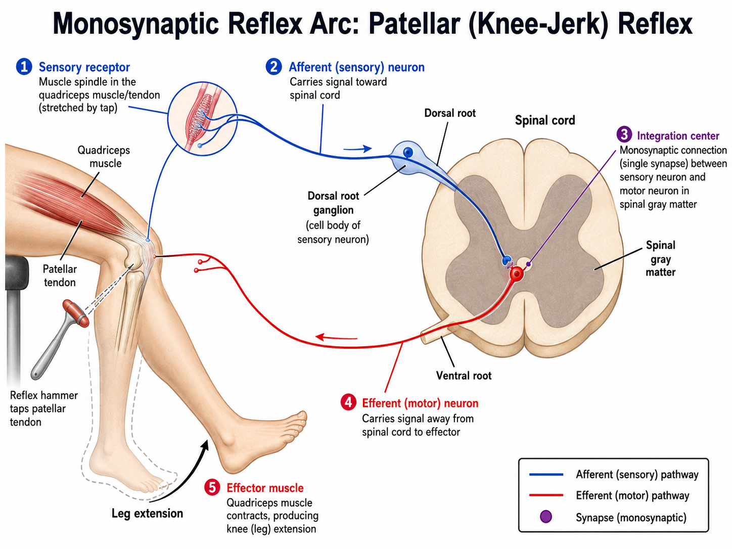
- Sensory receptor — detects the stimulus.
- Afferent (sensory) neuron — enters via the dorsal root (cell bodies in the dorsal root ganglion).
- Integration center — spinal cord or brainstem; interneurons process here.
- Efferent (motor) neuron — exits via the ventral root. Mnemonic: dorsal = afferent in, ventral = motor out.
- Effector — muscle or gland.
Monosynaptic vs. Polysynaptic Reflexes
Must knowKnow the distinction. The patellar (knee-jerk) reflex is the canonical monosynaptic reflex: the sensory neuron from the quadriceps muscle spindle synapses directly onto the motor neuron — no interneurons. Tapping the tendon stretches the quad → spindle fires → motor neuron contracts the quad → leg extends; reciprocal inhibition relaxes the antagonist hamstring. The muscle spindle detects stretch/length; contrast with the Golgi tendon organ, which detects tension and triggers the inverse stretch reflex (relax) to protect the tendon. Spindle = stretch → contract; Golgi tendon = tension → relax.
Polysynaptic reflexes involve interneurons. The withdrawal (flexor) reflex is the prime example: stepping on a nail → nociceptor → afferent → interneuron → motor neuron → flexor contracts (withdrawal), while the contralateral leg extends (crossed extensor reflex) to bear weight.
The Role of Supraspinal Circuits
Must knowThe spinal cord executes reflexes independently, but the brain continuously modulates them. Supraspinal circuits (cortex, brainstem, cerebellum) send descending pathways that inhibit or facilitate spinal reflexes, allowing voluntary override. After a spinal cord injury above the reflex level, cutting descending inhibitory pathways causes hyperreflexia (exaggerated reflexes).
The feedback loop underlying reflexes: stimulus → response → response removes/corrects the stimulus → feedback to the receptor. This is negative feedback, the same logic as thermoregulation and hormone control.
Quick check: A patient has a spinal cord transection at T10. Will she retain patellar reflexes (L3–L4)? Will she be able to voluntarily move her legs?
Yes, the patellar reflex is retained (and likely exaggerated, due to lost supraspinal inhibition) because the L3–L4 arc is intact below the injury. She will not voluntarily move her legs because descending motor pathways from the cortex are severed.
Integration with the Endocrine System: Feedback Control
The Nervous System and Endocrine System as Partners
Must knowThe nervous and endocrine systems are deeply integrated: fast, precise neural signaling complements sustained, broad hormonal action. Their meeting point is the hypothalamus.
The Hypothalamus–Pituitary Axis
Must knowThe hypothalamus translates neural input into hormonal output via two pathways to the pituitary gland:
- Hypothalamus → Anterior Pituitary: the hypothalamus releases releasing/inhibiting hormones into the hypothalamic-hypophyseal portal system (a direct portal vessel), which then stimulate or suppress anterior pituitary hormones (TSH, ACTH, LH, FSH, GH, prolactin).
- Hypothalamus → Posterior Pituitary: the posterior pituitary is neural tissue — an extension of the hypothalamus. Hypothalamic neurons synthesize ADH (vasopressin) and oxytocin and transport them down their axons for storage/release from the posterior pituitary (neurosecretion — neurons acting as endocrine cells).
Feedback Loops in the HPA (and Other) Axes
Must knowKnow the negative-feedback architecture. Example — the HPA axis:
Cortisol feeds back to inhibit both CRH and ACTH, keeping itself in range. The same three-tier pattern (hypothalamus → anterior pituitary → target gland → negative feedback) applies to the HPT (thyroid), HPG (gonads), and GH axes.
Neural Control of Endocrine Glands
Must knowBeyond the pituitary, the ANS directly controls several endocrine structures:
- Adrenal medulla: sympathetic preganglionic input → releases epinephrine, extending the fight-or-flight response beyond the acute neural signal.
- Pancreas: sympathetic input inhibits insulin/stimulates glucagon; parasympathetic stimulates insulin.
- Pineal gland: sympathetic light-dark signals → melatonin for circadian rhythms.
Quick check: A patient's adrenal cortex is destroyed (Addison's disease), eliminating cortisol production. What happens to CRH and ACTH levels, and why?
With no cortisol to feed back, both CRH and ACTH rise (negative feedback is absent). High ACTH (sharing a precursor with MSH) causes the characteristic skin hyperpigmentation.
Common Confusions & Tricks
1. Afferent vs. Efferent. Use SAME: Sensory = Afferent, Motor = Efferent. Afferent arrives at the CNS; efferent exits.
2. Sympathetic vs. Parasympathetic neurotransmitters. Both use ACh preganglionically (nicotinic). Sympathetic switches to NE postganglionically (adrenergic); parasympathetic stays ACh (muscarinic). The sweat-gland exception (sympathetic but muscarinic) is a classic trap — sympathetic does NOT always mean adrenergic.
3. "Craniosacral" vs. "Thoracolumbar." Parasympathetic = CrAnioSacral; Sympathetic = ThorAcoLumbar. The vagus nerve (CN X) is the giant of parasympathetic outflow.
4. Anterior vs. Posterior Pituitary. The posterior pituitary stores/releases hormones (ADH, oxytocin) MADE in the hypothalamus — it's neural tissue. The anterior pituitary makes its own hormones under hypothalamic control via portal blood. "Storing ≠ making."
5. Reflexes and consciousness. Reflexes do NOT require conscious awareness and complete before pain is perceived. Cortical awareness is a parallel, slower process.
6. Hyperreflexia after spinal cord injury. Cutting descending inhibitory pathways unleashes spinal reflexes → hyperreflexia and spasticity above the injury. If the reflex arc itself is destroyed, you get flaccid paralysis (no reflexes). Know the difference.
7. Myelin and cell type. CNS myelin → oligodendrocytes; PNS myelin → Schwann cells. MS attacks CNS myelin; Guillain-Barré attacks PNS myelin.
8. Acetylcholinesterase inhibitors → continuous ACh stimulation. Organophosphates, neostigmine, physostigmine prevent ACh breakdown → prolonged depolarization and desensitization → paralysis. Neostigmine treats myasthenia gravis; organophosphates are poison.
9. The adrenal medulla releases epinephrine (mostly), not just NE. Epinephrine has stronger β₂ effects (bronchodilation, muscle vasodilation) — useful for drug-mechanism questions.
Key Takeaways
Organization
- CNS = brain + spinal cord (integration); PNS = all nerves outside CNS (sensory in + motor out)
- PNS divides into somatic (voluntary, single neuron) and autonomic (involuntary, two-neuron chain)
- ANS divides into sympathetic (thoracolumbar, fight-or-flight) and parasympathetic (craniosacral, rest-and-digest)
Neurons and Signaling
- Three types: sensory (afferent), interneurons (within CNS), motor (efferent)
- Myelin (oligodendrocytes CNS, Schwann cells PNS) → saltatory conduction → faster signals
- Action potential: all-or-nothing; −70 mV rest → −55 mV threshold → depolarization → repolarization → hyperpolarization; absolute then relative refractory period
- Synaptic transmission: Ca²⁺ triggers vesicle fusion → NT release → EPSP or IPSP → summation at axon hillock
- Key NTs: ACh (NMJ, ANS), NE (sympathetic post), dopamine (reward/movement), GABA (inhibition), glutamate (excitation), serotonin (mood)
Autonomic Nervous System
- Both divisions: preganglionic ACh (nicotinic)
- Parasympathetic postganglion: ACh (muscarinic); Sympathetic postganglion: NE (adrenergic) — except sweat glands (ACh)
- Adrenal medulla = modified sympathetic ganglion → epinephrine into blood
Reflexes
- Reflex arc: receptor → afferent → integration (spinal cord) → efferent → effector
- Monosynaptic: patellar (L3–L4), no interneurons; Polysynaptic: withdrawal/flexor + crossed extensor
- Supraspinal circuits modulate reflexes; cutting descending paths → hyperreflexia
Neuroendocrine Integration
- Hypothalamus = master interface between nervous and endocrine systems
- Anterior pituitary controlled via hypothalamic-hypophyseal portal system; posterior pituitary stores/releases ADH and oxytocin made in hypothalamus (neurosecretion)
- All major axes (HPA, HPT, HPG) run on three-tier negative feedback
- Sympathetic → adrenal medulla → epinephrine: neural signal extended as hormonal signal