The circulatory system is one of the highest-yield MCAT topics — it appears in cardiac, renal, and respiratory passage contexts. Your goal is to understand it as an integrated pressure-and-flow network, not a list of facts. Once you have that mental model, the details click into place.
Priority labels: Must know = cold; Know the logic = mechanism not names; Passage-level = recognize, don't memorize; Optional = skippable.
Functions of the Circulatory System
Must knowThe circulatory system is a delivery and disposal service operating under pressure. Its core functions:
- Oxygen delivery and removal
- Nutrient transport (glucose, amino acids, fatty acids)
- Hormone distribution from glands to targets
- Ion and fluid balance (with the kidneys)
- Waste removal (urea, creatinine, lactic acid)
- Immune function — WBCs patrol via blood
- Thermoregulation — blood redistributes heat
Blood is a multipurpose courier. The MCAT often asks you to connect a circulatory disruption (e.g., low cardiac output) to downstream consequences in multiple organ systems at once.
Role in Thermoregulation
Must knowBlood is a good heat-transfer medium (high specific heat). Heat from the core is carried to the skin and dissipated. Vasodilation of cutaneous vessels increases skin flow and heat loss (why you flush when hot); vasoconstriction shunts blood away from the periphery to conserve heat.
Passage-levelCountercurrent heat exchange: in the limbs, warm arterial blood transfers heat to cooler venous blood returning to the core before it's lost to the environment, conserving body heat (same principle as fish gills).
Quick check: If a patient has a fever and the body is trying to dissipate heat, would peripheral vessels dilate or constrict? Answer: Dilate — vasodilation increases surface flow to radiate excess heat (why people look flushed during fever).
The Four-Chambered Heart: Structure and Function
Anatomy and Valves
Must knowThe heart has four chambers: two atria (thin-walled, receiving) and two ventricles (thick-walled, pumping). The right side receives deoxygenated blood and pumps it to the lungs; the left side receives oxygenated blood and pumps it to the body. The left ventricle has the thickest wall (highest pressure). The septum prevents mixing of oxygenated and deoxygenated blood.
The valves prevent backflow:
- Atrioventricular (AV) valves (between atria and ventricles): tricuspid (3 cusps) on the right, bicuspid/mitral on the left. Close when ventricles contract → heart sound S1 ("lub").
- Semilunar valves (ventricle exits): pulmonary (right), aortic (left). Close at the end of systole → heart sound S2 ("dub").
Mnemonic: "Try Puking" (Tricuspid, Pulmonary) on the right.
Cardiac Conduction System
Must knowThe heart is autorhythmic (generates its own impulses). The sequence:
- Sinoatrial (SA) node — the pacemaker, in the right atrium; sets resting HR (~60–100 bpm).
- Signal spreads across the atria → atria contract.
- Atrioventricular (AV) node — adds a brief delay so atria empty before ventricles contract.
- Bundle of His → bundle branches → Purkinje fibers → ventricles contract from the apex upward, wringing blood into the great vessels.
Sympathetic stimulation speeds SA depolarization (faster HR); parasympathetic (vagal) ACh slows it.
The Cardiac Cycle
Must know- Systole — ventricular contraction; AV valves closed, semilunar open; blood ejected.
- Diastole — ventricular relaxation and filling; AV valves open, semilunar closed.
Cardiac output (CO) is volume pumped per minute:
where is stroke volume (~70 mL/beat at rest, giving CO ≈ 5 L/min).
Know the logicThree factors govern SV:
- Preload — ventricular stretch at end-diastole; more stretch → stronger contraction (Frank-Starling law: the heart pumps whatever it receives, automatically matching right and left output).
- Afterload — resistance the ventricle ejects against (arterial pressure); higher afterload → lower SV.
- Contractility — intrinsic contraction force; sympathetic/epinephrine increase it (positive inotropy).
Quick check: A patient hemorrhages. Trace the effect on preload, SV, and CO. Answer: Less blood volume → less venous return → lower preload → less stretch → lower SV → lower CO. The body compensates with increased HR (sympathetic activation).
Endothelial Cells
Must knowEndothelial cells line the entire interior of the cardiovascular system, forming a continuous, metabolically active monolayer (the endothelium) — not just a passive barrier.
Know the logicKey functions:
- Nitric oxide (NO) — diffuses into smooth muscle causing relaxation/vasodilation; also inhibits platelet aggregation. Loss of endothelial function (atherosclerosis) reduces NO → vasoconstriction and clotting.
- Selective permeability — regulate what crosses between blood and tissue (e.g., blood-brain barrier tight junctions).
- Optional also release prostacyclin (vasodilator) and endothelin (vasoconstrictor); drive angiogenesis.
Quick check: Why does atherosclerosis increase hypertension risk? Answer: Plaque damages endothelial cells → less NO → impaired vasodilation + vessel stiffening → increased peripheral resistance → elevated BP.
Systolic and Diastolic Pressure
Must knowBlood pressure (BP) is reported as systolic/diastolic in mmHg; normal ≈ 120/80.
- Systolic (~120): peak pressure during ventricular contraction
- Diastolic (~80): minimum pressure during relaxation
- Pulse pressure = systolic − diastolic
Mean arterial pressure (MAP) is the average driving force for tissue perfusion. Because diastole lasts ~twice as long as systole:
MAP also relates to cardiac output and resistance (analogous to Ohm's law, ):
where is total peripheral resistance. You can raise MAP by increasing CO or resistance (vasoconstriction).
Quick check: If a drug doubles TPR while CO stays constant, what happens to MAP? Answer: MAP doubles (MAP = CO × TPR) — models hypertension from excessive vasoconstriction.
Pulmonary and Systemic Circulation
Must knowThe system has two circuits in series:
Pulmonary Circulation
Must knowRight ventricle → pulmonary artery → pulmonary capillaries (gas exchange) → pulmonary veins → left atrium
- Operates at low pressure; the right ventricle is thinner because pulmonary resistance is low.
- Counterintuitive naming: pulmonary arteries carry deoxygenated blood; pulmonary veins carry oxygenated blood. "Artery/vein" refers to direction relative to the heart, NOT oxygen content.
Systemic Circulation
Must knowLeft ventricle → aorta → arteries → arterioles → capillaries → venules → veins → vena cavae → right atrium
- Operates at high pressure (MAP ≈ 93 mmHg); the left ventricle is the thickest chamber.
Since the circuits are in series, right and left ventricular output must be equal over time.
Portal Systems
Passage-levelA portal system sends blood through two capillary beds in series before returning to the heart. The main example is the hepatic portal system: nutrient-rich GI blood drains via the hepatic portal vein to the liver for processing before reaching the inferior vena cava. (Also: hypothalamic-hypophyseal portal system to the anterior pituitary.)
Fetal Circulation
Know the logicFetal lungs are nonfunctional, so blood bypasses the lungs via three shunts. Oxygenated blood arrives from the placenta via the umbilical vein:
- Ductus venosus — umbilical vein past the liver to the IVC.
- Foramen ovale — right atrium directly to left atrium, bypassing the lungs.
- Ductus arteriosus — pulmonary artery to aorta, bypassing the lungs.
Deoxygenated blood returns via the umbilical arteries. At birth, the first breath drops pulmonary resistance; the foramen ovale and ductus arteriosus close.
Quick check: A patient has a patent foramen ovale. Given left atrial pressure > right, which way does blood shunt, and what's the consequence? Answer: Left (oxygenated) → right atrium, back to the lungs — a left-to-right shunt; oxygenated blood is "wasted." If severe, can progress to pulmonary hypertension and reverse to a right-to-left shunt with cyanosis (Eisenmenger syndrome).
Arterial and Venous Systems
Vessel Types and Structure
Must knowAll vessels except capillaries have three layers: tunica intima (endothelium), tunica media (smooth muscle + elastic tissue, controls diameter), and tunica adventitia (connective tissue). The tunica media is thickest in arteries, letting them withstand pressure and recoil.
Arteries and Arterioles
Must know- Elastic arteries (aorta, large arteries) expand in systole and recoil in diastole, maintaining continuous flow (why diastolic pressure isn't zero).
- Muscular arteries distribute blood to specific regions.
- Arterioles are the primary site of peripheral resistance: their thick smooth muscle constricts/dilates dramatically, acting as the main "faucet" for flow to capillary beds.
Capillaries
Must knowCapillaries are the units of exchange — a single endothelial layer (~1 cell thick), no media or adventitia. Features that optimize exchange: tiny diameter (RBCs deform to pass), ~1 μm wall (short diffusion distance), and an enormous total cross-sectional area → very slow flow → maximum contact time.
Venules and Veins
Must knowVeins have thinner walls, larger lumens, and less smooth muscle than arteries; they operate at low pressure. About 60–70% of blood volume is stored in veins — the capacitance (reservoir) vessels, mobilized by venoconstriction. Veins have one-way valves; the skeletal muscle pump and respiratory pump aid venous return against gravity. (Venules are also where WBCs exit to tissue.)
Pressure and Flow Through the Circulation
Must knowFlow follows Poiseuille's law:
where = flow, = pressure gradient, = radius, = viscosity, = length. The critical insight is the fourth-power dependence on radius: doubling radius increases flow 16-fold — why small arteriolar diameter changes hugely alter tissue flow. Resistance is .
Moving aorta → arterioles → capillaries, individual radius decreases but total cross-sectional area increases dramatically. Therefore:
- Velocity is inversely proportional to total cross-sectional area — slowest in capillaries, fastest in the aorta.
- Pressure drops progressively, with the steepest drop across arterioles (greatest resistance).
Quick check: Why does blood flow slowly in capillaries despite high cardiac pressure? Answer: The combined cross-sectional area of all capillaries vastly exceeds the aorta. Since flow is conserved, velocity drops proportionally — slow flow = more time for exchange.
Capillary Beds: Exchange Mechanisms
Mechanisms of Gas and Solute Exchange
Must knowGases (, ) and small lipid-soluble molecules cross by simple diffusion down concentration gradients. Larger water-soluble molecules use fenestrations (in fenestrated capillaries) or transcytosis, plus bulk flow.
Starling Forces: Filtration and Reabsorption
Must knowFluid movement across capillary walls is the balance of hydrostatic (pushes fluid out) and oncotic (pulls fluid in, mainly from plasma albumin) pressures — the Starling forces:
At the arterial end, hydrostatic pressure dominates → net filtration out. At the venous end, oncotic pressure dominates → net reabsorption in. The small net excess of filtrate is returned by the lymphatic system.
Edema results from disrupting this balance: low plasma protein (cirrhosis, nephrotic syndrome) → low oncotic pressure → excess filtration; high venous pressure (heart failure) → reduced reabsorption.
Source of Peripheral Resistance
Must knowPeripheral resistance arises primarily from the arterioles (abundant smooth muscle → large diameter changes). Other contributors: blood viscosity (↑ in polycythemia, ↓ in anemia) and vessel length. Increasing resistance raises arterial pressure; vasodilation lowers it.
Quick check: In anemia, blood viscosity decreases. Per Poiseuille's law, how does this affect flow? Answer: Lower viscosity = lower resistance = higher flow for the same gradient. This partly explains the high cardiac output of anemic patients (compensating for reduced O₂-carrying capacity).
Composition of Blood
Must knowSpinning a blood sample separates:
- Plasma (~55%) — the liquid portion
- Buffy coat — WBCs and platelets
- Erythrocytes (RBCs) — the dense pellet
Plasma
Must knowPlasma is ~90% water and contains plasma proteins (albumin — oncotic pressure and transport; globulins — antibodies; fibrinogen — clotting), electrolytes (Na⁺, K⁺, Ca²⁺, Cl⁻, HCO₃⁻), nutrients, wastes (urea, creatinine, bilirubin), and hormones.
Serum = plasma minus clotting factors.
Blood Cells
Must know| Cell Type | Function |
|---|---|
| Erythrocytes (RBCs) | O₂/CO₂ transport |
| Leukocytes (WBCs) | Immunity |
| Platelets | Hemostasis |
All are produced in bone marrow.
Erythrocytes: Production and Destruction
Must knowErythropoiesis occurs in red bone marrow. The hormone erythropoietin (EPO), secreted by the kidney in response to hypoxia, accelerates RBC production (basis for EPO doping and treating CKD anemia).
During maturation, RBCs eject their nucleus and organelles — mature RBCs are anucleate, can't synthesize proteins, and rely on anaerobic glycolysis for ATP. Their biconcave shape maximizes surface-area-to-volume and lets them deform through capillaries.
RBC lifespan is ~120 days. Old RBCs are removed by macrophages, mainly in the spleen (and liver). Hemoglobin breakdown: globin → amino acids; heme → bilirubin (excreted in bile); iron → recycled.
Quick check: A patient with chronic kidney disease develops anemia. Explain. Answer: Damaged kidneys produce insufficient EPO → less marrow stimulation → fewer RBCs → anemia. Treated with recombinant EPO.
ABO and Rh Blood Groups
Passage-levelRBC surface antigens define blood type; plasma antibodies against the absent antigens determine compatibility.
| Type | Antigen | Antibodies | Receive from |
|---|---|---|---|
| A | A | anti-B | A, O |
| B | B | anti-A | B, O |
| AB | A, B | none | all (universal recipient) |
| O | none | anti-A, anti-B | O only (universal donor) |
Incompatible blood → antibodies bind donor antigens → agglutination and hemolysis.
The Rh factor (D antigen) is separate: Rh⁻ individuals make anti-D only after exposure. This underlies hemolytic disease of the newborn (Rh⁻ mother sensitized to a first Rh⁺ fetus makes anti-Rh IgG attacking a later Rh⁺ fetus; prevented by RhoGAM).
Regulation of Plasma Volume
Must knowPlasma volume sets venous return, preload, and cardiac output. Hormonal control:
- RAAS: low BP/flow → kidney releases renin → angiotensinogen → angiotensin I → ACE converts to angiotensin II (vasoconstriction) + stimulates aldosterone → kidneys retain Na⁺ (water follows) → volume up.
- ADH (vasopressin): released from posterior pituitary when plasma osmolarity rises or volume falls → increases water reabsorption in the collecting duct → volume up.
- Atrial natriuretic peptide (ANP): released when atria are stretched (high volume) → promotes Na⁺/water excretion → volume down (opposes RAAS/ADH).
Quick check: A patient takes an ACE inhibitor. Predict effects on aldosterone and BP. Answer: Less angiotensin II → less aldosterone → more Na⁺/water lost → lower volume, plus less angiotensin II–mediated vasoconstriction → BP decreases. ACE inhibitors are a cornerstone of hypertension/heart-failure treatment.
Coagulation and Clotting Mechanisms
Must knowHemostasis stops bleeding in three overlapping stages:
- Vascular spasm — immediate reflexive vasoconstriction at the injury.
- Platelet plug (primary hemostasis) — exposed collagen (with von Willebrand factor) binds platelets, which activate (releasing ADP, thromboxane A₂) and aggregate via fibrinogen into a loose plug.
- Coagulation cascade (secondary hemostasis) — plasma clotting factors reinforce the plug. Know the logic The common pathway activates prothrombin → thrombin, and thrombin converts fibrinogen → fibrin, which polymerizes into an insoluble mesh stabilizing the clot.
Fibrinolysis: plasminogen → plasmin (via tPA) degrades fibrin once the vessel heals.
Oxygen Transport by Blood
Hemoglobin
Must knowHemoglobin (Hb) is a tetramer of 4 globin chains, each with a heme group containing Fe²⁺ that binds one . Adult HbA has two α and two β subunits.
Hematocrit is the percentage of blood volume that is RBCs (~42–47% males, ~37–42% females) — a proxy for oxygen-carrying capacity.
OptionalO₂ content has two parts — bound to Hb () plus a negligible dissolved fraction.
The Oxyhemoglobin Dissociation Curve
Must knowHemoglobin shows cooperative binding — one binding raises affinity of the rest — producing the sigmoidal vs curve.
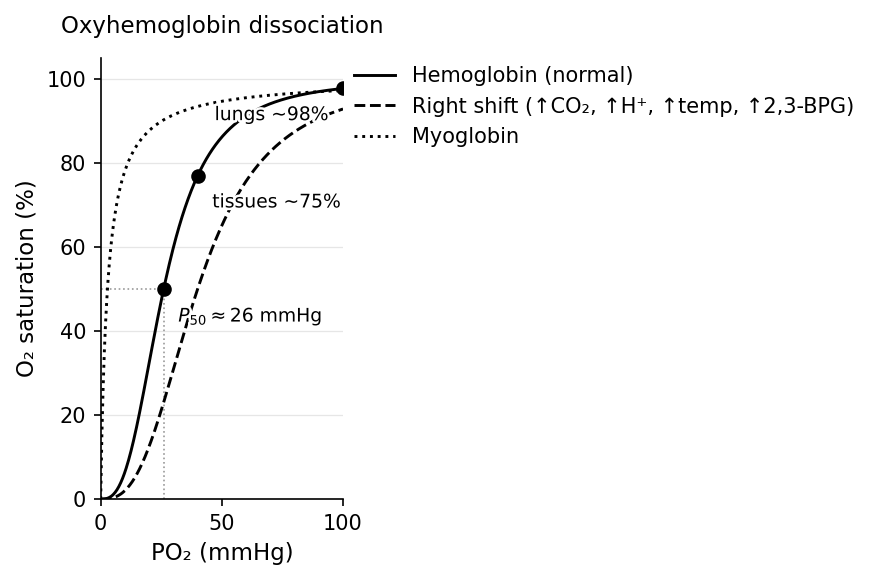
Contrast myoglobin (monomeric muscle O₂-storage protein), which has a hyperbolic curve and higher O₂ affinity than Hb — so it extracts O₂ from Hb in muscle.
Key points: ~98% saturated in the lungs ( ≈ 100), ~75% in resting tissue (≈ 40), dropping much lower in exercising tissue. The P₅₀ (the for 50% saturation, ~26 mmHg) measures affinity: higher P₅₀ = lower affinity = right shift.
Factors Shifting the Curve (Bohr Effect)
Must knowA right shift (lower affinity) promotes O₂ unloading to tissues.
| Factor | Right shift (↓ affinity) |
|---|---|
| pH | Acidic (↓ pH) |
| CO₂ | High |
| Temperature | High |
| 2,3-BPG | High |
The Bohr effect is the pH/CO₂ piece: active tissues produce , lowering pH, right-shifting the curve, and releasing O₂ where it's needed; in the lungs the reverse promotes loading.
Know the logic- 2,3-BPG (a glycolysis byproduct in RBCs) stabilizes deoxyhemoglobin → right shift. High altitude → ↑2,3-BPG → enhanced O₂ delivery (acclimatization).
- Fetal hemoglobin (HbF) binds 2,3-BPG poorly → left-shifted, higher affinity → extracts O₂ from maternal blood across the placenta.
- Carbon monoxide (CO) binds Fe²⁺ ~250× more tightly than O₂, forming carboxyhemoglobin and left-shifting the remaining Hb — a double harm.
Quick check: Compare a climber's O₂ curve at sea level vs. after 3 weeks at altitude, and explain the value. Answer: ↑2,3-BPG right-shifts the curve → at any , Hb unloads more O₂, so tissues stay supplied despite lower arterial . EPO also raises RBC count, further improving delivery.
Carbon Dioxide Transport and Levels in Blood
Must knowfrom tissue metabolism is transported to the lungs in three forms:
| Form | Proportion |
|---|---|
| As bicarbonate () | ~70% |
| Carbaminohemoglobin (bound to globin) | ~20–23% |
| Dissolved in plasma | ~7–10% |
Bicarbonate Transport (Most Important)
Must knowInside RBCs, reacts with water via carbonic anhydrase:
The exits the RBC via the chloride shift ( out, in). The is buffered by deoxyhemoglobin, which carries CO₂/H⁺ better than oxyhemoglobin (Haldane effect). At the lungs the reaction reverses and is exhaled.
Carbaminohemoglobin
Must knowbinds the amino groups of globin (not heme iron); deoxyhemoglobin binds it more readily (Haldane effect again).
Quick check: Hyperventilation lowers blood . What happens to pH? Answer: Less CO₂ drives in reverse → less H⁺ → pH rises (respiratory alkalosis), which can cause tingling and cramps.
Nervous and Endocrine Control of the Circulatory System
Autonomic Nervous Control
Must knowSympathetic ("fight or flight"): releases norepinephrine (and adrenal epinephrine) → ↑ heart rate (positive chronotropy) and contractility (positive inotropy) via β₁ → ↑ CO; vasoconstriction in most vessels (α₁), vasodilation in skeletal muscle/coronary (β₂). Net: ↑ CO and TPR → ↑ BP, blood redirected to muscles.
Parasympathetic (vagus, CN X): releases acetylcholine → slows heart rate (negative chronotropy) via the SA node; minimal effect on most systemic vessels.
Baroreceptor Reflex
Must knowBaroreceptors are stretch receptors in the carotid sinus and aortic arch that fire in proportion to arterial pressure — a fast negative feedback loop:
- BP rises → fire more → medulla ↑ parasympathetic / ↓ sympathetic → HR slows, vasodilation → BP falls toward normal.
- BP drops → fire less → ↓ parasympathetic / ↑ sympathetic → HR and vasoconstriction increase → BP rises.
This explains orthostatic hypotension (gravity pools blood on standing; baroreceptors trigger compensatory tachycardia) and the blunted HR response on beta-blockers.
Chemoreceptors
Passage-levelPeripheral chemoreceptors (carotid/aortic bodies) respond to low , high , low pH → ↑ ventilation + sympathetic vasoconstriction. Central chemoreceptors (medulla) respond mainly to /pH and are the dominant ventilatory drive.
Endocrine Control
Must know| Hormone | Source | Effect |
|---|---|---|
| Epinephrine | Adrenal medulla | ↑ HR, ↑ contractility (β₁) |
| Angiotensin II | Renin-ACE cascade | Vasoconstriction; ↑ aldosterone |
| Aldosterone | Adrenal cortex | ↑ Na⁺/H₂O retention → ↑ BP |
| ADH | Posterior pituitary | ↑ H₂O retention; vasoconstriction |
| ANP | Atrial cardiomyocytes | ↓ Na⁺/H₂O → ↓ volume |
| EPO | Kidney | ↑ RBC production |
Quick check: A patient in hypovolemic shock has low BP. List two fast nervous and two slower hormonal responses. Answer: Fast: (1) baroreceptors → ↑ sympathetic → tachycardia + vasoconstriction; (2) ↑ NE/Epi → more vasoconstriction and contractility. Slow: (1) RAAS → angiotensin II vasoconstriction + aldosterone Na⁺ retention; (2) ADH → water retention + vasoconstriction.
Common Confusions & Tricks
1. Arteries vs. veins: direction, not oxygen content.
Pulmonary arteries carry deoxygenated blood; pulmonary veins carry oxygenated blood. Artery = away from heart; vein = toward heart.
2. Hemoglobin binding curve vs. Michaelis-Menten kinetics.
The Hb–O₂ curve (sigmoidal) describes cooperative ligand binding, not enzyme kinetics. Don't call it Michaelis-Menten. Myoglobin's hyperbolic curve reflects simple binding. Michaelis-Menten describes enzyme catalysis — a different phenomenon.
3. Left shift vs. right shift: which is "good"?
Neither universally. A right shift helps unload O₂ in active tissues; a left shift helps load O₂ where is low (fetal Hb, lungs). Context decides.
4. Systolic vs. diastolic.
Systolic = contraction = higher number = semilunar valves open, AV closed. Diastolic = relaxation = lower number = AV open. S1 = AV closure (start of systole); S2 = semilunar closure (end of systole).
5. Serum vs. plasma.
Plasma = liquid portion (with clotting factors). Serum = plasma minus clotting factors. Albumin is in both; fibrinogen only in plasma.
6. Right ventricle has low pressure — not "less important."
The lower pressure suits the low-resistance pulmonary circuit. Right heart failure → systemic (peripheral) edema; left heart failure → pulmonary edema.
7. CO poisoning = two problems.
Less O₂ can bind (CO occupies sites) AND remaining Hb is left-shifted (holds O₂ too tightly). Treatment: 100% O₂ or hyperbaric oxygen.
8. Peripheral resistance lives in arterioles, not arteries (conduits) or capillaries (exchange).
9. Hemostasis has three sequential stages.
Vascular spasm → platelet plug (primary) → coagulation cascade producing a fibrin mesh (secondary). The fibrin comes from thrombin cleaving fibrinogen.
10. 2,3-BPG: HbF vs. HbA.
HbF's γ-subunits bind 2,3-BPG poorly → HbF is NOT right-shifted → stays left-shifted relative to HbA → extracts O₂ from maternal blood.
11. ANP opposes RAAS/ADH.
High volume → atria stretch → ANP → natriuresis/diuresis → volume down. Low volume → RAAS + ADH → Na⁺/H₂O retention → volume up. The MCAT loves asking what happens when one system is blocked.
Key Takeaways
The Big Picture
- The circulation is a pressure-driven, resistance-regulated network (MAP = CO × TPR) with two circuits in series (pulmonary and systemic), driven by the heart and tuned mainly at the arterioles.
- Arteries carry blood away from the heart, veins toward it — direction, not oxygen content (pulmonary artery is deoxygenated; pulmonary veins oxygenated).
- Capillaries are the only exchange site; Starling forces drive filtration at the arterial end and reabsorption at the venous end, with lymphatics returning the excess.
- Blood is plasma (~55%) plus formed elements; RBCs carry O₂, WBCs provide immunity, platelets initiate hemostasis (vascular spasm → platelet plug → cascade ending in a thrombin-generated fibrin mesh).
- Oxygen rides on hemoglobin with a sigmoidal, cooperative curve; right shifts (↑CO₂, ↑H⁺, ↑temp, ↑2,3-BPG — the Bohr effect) promote unloading. Most CO₂ travels as bicarbonate via carbonic anhydrase.
- BP is regulated fast by the baroreceptor reflex and longer-term by hormonal volume control (RAAS, ADH raise volume; ANP lowers it).