The MCAT tests the immune system deeply — expect passages that ask you to reason through unfamiliar immune scenarios using solid conceptual foundations. This guide builds from the fast, non-specific first responders to the highly specific adaptive machinery that remembers a pathogen for decades.
Priority labels: Must know = cold; Know the logic = mechanism not names; Passage-level = recognize, don't memorize; Optional = skippable.
Overview: Two Layers of Defense
Must knowInnate (nonspecific) immunity is immediate (minutes to hours), does not improve with exposure, and recognizes broad molecular patterns rather than specific pathogens. Adaptive (specific) immunity is slower on first exposure (days to weeks) but highly specific, improves on reexposure, and generates immunological memory.
Key Distinctions
Must know| Feature | Innate | Adaptive |
|---|---|---|
| Speed | Immediate (minutes–hours) | Delayed (days–weeks) |
| Specificity | Broad (pattern recognition) | Exquisitely specific (one antigen) |
| Memory | None | Yes — memory cells formed |
| Cells | Neutrophils, macrophages, NK cells, dendritic cells, mast cells | T lymphocytes, B lymphocytes |
| Receptors | Pattern recognition receptors (PRRs), e.g., Toll-like receptors (TLRs) | T-cell receptors (TCRs), B-cell receptors (BCRs) |
The two work together: innate immunity buys time, drives inflammation, and — critically — activates the adaptive response by presenting antigen to lymphocytes.
Quick check: A patient with a mutation that completely disables Toll-like receptors would have impaired function in which arm of immunity, and what specific consequence would you expect?
Answer: Innate immunity. TLRs are pattern recognition receptors that detect conserved microbial structures (LPS, flagellin, viral dsRNA). Loss of TLRs would blunt the early inflammatory response and impair activation of the adaptive response, because innate signaling provides critical co-stimulatory signals to T cells.
Innate Immune System Cells
Must knowInnate cells are "pre-armed" — no clonal expansion or receptor recombination needed. They recognize pathogen-associated molecular patterns (PAMPs) on microbes via pattern recognition receptors.
Neutrophils
Must knowNeutrophils are the most abundant WBCs and the first responders to infection. They are aggressively phagocytic, killing bacteria with reactive oxygen species (the oxidative burst) and granule enzymes, and can release neutrophil extracellular traps (NETs) to trap pathogens. They are short-lived and characteristic of acute bacterial infection — high neutrophil count on a CBC should suggest bacterial infection.
Macrophages
Must knowMacrophages are the workhorses of innate immunity and a key bridge to adaptive immunity. They derive from circulating monocytes. Three key functions:
- Phagocytosis — engulf and destroy pathogens, dead cells, debris.
- Antigen presentation — display digested fragments on MHC class II to activate helper T cells.
- Cytokine secretion — release pro-inflammatory cytokines that orchestrate the response and trigger fever.
Tissue-resident macrophages have special names — Kupffer cells (liver), microglia (brain), osteoclasts (bone), alveolar macrophages (lungs), Langerhans cells (skin).
Other Innate Cells
Must knowNatural killer (NK) cells are innate lymphocytes that do NOT use rearranged antigen-specific receptors. They surveil for cells that have downregulated MHC class I (a trick viruses and cancer cells use to hide from cytotoxic T cells) and kill them via perforin/granzyme. The rule: have MHC I → NK cells leave you alone; lack MHC I → NK cells kill you.
Dendritic cells are the premier antigen-presenting cells (APCs). They sample peripheral tissues, engulf pathogens, then migrate to lymph nodes to present antigen to naïve T cells — the most potent activators of the adaptive response.
Mast cells reside in tissues and, when activated via IgE during allergic reactions, release histamine (vasodilation, increased permeability) and other mediators. They are central to type I hypersensitivity (allergy/anaphylaxis). Basophils are their circulating counterparts; eosinophils target parasites and mediate late-phase allergy.
Quick check: A patient stung by a bee immediately develops hives, difficulty breathing, and low blood pressure. Which innate immune cell is primarily responsible, and through what mechanism?
Answer: Mast cells (and basophils). On first exposure, IgE coats mast cells; on second exposure, venom cross-links the IgE, triggering degranulation — rapid release of histamine and other mediators. This is anaphylaxis (type I hypersensitivity), where IgE from the adaptive arm arms the innate mast cells.
Innate Defenses: Barriers, Complement, Inflammation, and Interferons
First Line: Physical and Chemical Barriers
Know the logicSkin (tough, dry barrier), mucous membranes / mucociliary escalator (trap and sweep particles), lysozyme (cleaves bacterial peptidoglycan), low stomach pH, defensins (antimicrobial peptides), and normal flora (outcompete pathogens).
The Complement System
Must knowComplement is a group of plasma proteins (liver-made) that, once triggered by microbial surfaces or bound antibody, amplify innate defense. Know the three functional outcomes:
- Opsonization — coat microbes to tag them for phagocytosis.
- Inflammation — fragments recruit and activate immune cells.
- Lysis — terminal proteins form the membrane attack complex (MAC), which punches pores and lyses the target.
Out of scope: the factor-by-factor cascade (C1–C9, specific convertases) — focus on the three outcomes.
Inflammation
Know the logicInflammation produces four cardinal signs: redness, heat, swelling, pain.
- Damaged cells and mast cells release histamine, prostaglandins, cytokines → vasodilation (redness, heat) and increased vascular permeability (swelling/edema).
- Endothelium upregulates adhesion molecules → leukocytes undergo margination and rolling, then firm adhesion.
- Leukocytes squeeze between endothelial cells (diapedesis/extravasation) and follow chemotactic gradients to the infection site (chemotaxis).
Interferons
Know the logicInterferons are antiviral cytokines secreted by virus-infected cells that induce an antiviral state in neighboring cells, limiting viral spread before adaptive immunity engages.
Adaptive Immune System Cells: T Lymphocytes
Must knowAll lymphocytes originate from lymphoid progenitors in the bone marrow. T cells then migrate to the thymus to mature (T = thymus-derived), where they undergo selection and emerge as naïve T cells.
T cells are defined by their T-cell receptor (TCR), which can ONLY recognize antigen presented on an MHC molecule — never free, soluble antigen. This is MHC restriction.
Helper T Cells (CD4+)
Must knowHelper T cells express CD4 and recognize antigen on MHC class II (presented by APCs). They are the "commanders" — rather than killing directly, they secrete cytokines that:
- Activate cytotoxic T cells
- Provide co-stimulation to B cells (T cell-dependent B cell activation)
- Activate macrophages
Helper T cells differentiate into subsets tuned to the pathogen (intracellular vs. extracellular/parasitic). Regulatory T cells (Tregs) are a distinct subset that suppresses responses and maintains self-tolerance.
HIV preferentially infects CD4+ T cells (via the CD4 receptor); as counts fall, the patient progresses to AIDS.
Cytotoxic T Cells (CD8+)
Must knowCytotoxic T lymphocytes (CTLs) express CD8 and recognize antigen on MHC class I (on virtually all nucleated cells). When a CTL detects foreign peptide (viral or cancer) on MHC I, it kills the cell — primarily via perforin (pores) and granzymes (proteases that trigger apoptosis); Fas–FasL is an additional route.
Memory T Cells
Must knowAfter a response, most effector T cells die (contraction phase), but a subset persists as long-lived memory T cells that respond faster and more robustly on re-exposure — the cellular basis of immunological memory.
Quick check: A person's CD4+ helper T cell count is severely depleted (as in AIDS). What happens to their ability to make antibodies against a new pathogen, and why?
Answer: Antibody production is severely impaired. Most B cell activation is T cell-dependent — B cells need cytokine signals from CD4+ helper T cells to proliferate, class-switch, and become plasma cells. Without helper T cells, they cannot make high-affinity class-switched antibody, which is why AIDS patients are vulnerable to opportunistic infections.
Adaptive Immune System Cells: B Lymphocytes
Must knowB lymphocytes mature in the bone marrow in humans. Their defining feature is the B-cell receptor (BCR) — a membrane-bound antibody. Unlike T cells, B cells CAN recognize native, free, soluble antigen directly — no MHC required.
When activated (usually needing both BCR engagement AND helper T cell signals), a B cell proliferates and differentiates into:
- Plasma cells — terminally differentiated antibody factories that have lost their surface BCR and secrete massive amounts of antibody.
- Memory B cells — long-lived cells that respond rapidly on re-exposure and produce high-affinity antibodies (having undergone somatic hypermutation and affinity maturation).
Class Switching (Isotype Switching)
Know the logicB cells initially make IgM, then — under helper T cell cytokine signals — switch to other classes (IgG, IgA, IgE). The variable region (antigen specificity) does NOT change — only the constant region (and thus function) does.
Quick check: A patient has a genetic defect preventing class switching. What antibody class would they always produce, and what would be missing?
Answer: Only IgM. They would lack IgG (opsonization, complement, placental transfer), IgA (mucosal immunity), and IgE (parasitic defense, allergy) — similar to Hyper-IgM syndrome.
Tissues of the Immune System
Must knowPrimary lymphoid organs = where lymphocytes develop and mature. Secondary lymphoid organs = where immune responses are initiated.
Bone Marrow (Primary)
Must knowSite of hematopoiesis — all blood cells, including immune cells, arise here from hematopoietic stem cells. Both T and B cells originate here; B cells also complete maturation here. It is the long-term home of plasma and memory cells. Damage (radiation, chemo) devastates the whole immune system.
Thymus (Primary)
Must knowSite of T cell maturation via a two-step selection:
- Positive selection (thymic cortex): T cells that can recognize self MHC survive; those that cannot die by neglect. Ensures MHC restriction.
- Negative selection (medulla): T cells that bind self-MHC + self-peptide too strongly are deleted, eliminating autoreactive cells.
Result: self-tolerant, MHC-restricted naïve T cells exit. This is central tolerance; failure of negative selection contributes to autoimmunity.
Lymph Nodes (Secondary)
Must knowBean-shaped organs along lymphatic vessels that filter lymph and are primary sites where adaptive responses initiate. Optional anatomy: cortex (B cell follicles), paracortex (T cell zone), medulla (macrophages, plasma cells). Local nodes swell (lymphadenopathy) during clonal expansion.
Spleen (Secondary)
Must knowLargest secondary lymphoid organ; filters blood (vs. lymph nodes' lymph). Red pulp filters old/damaged RBCs; white pulp mounts responses to blood-borne antigens. Passage-level splenectomy patients are especially susceptible to encapsulated bacteria, because the spleen is the main site for opsonizing antibodies against polysaccharide capsules.
Quick check: If a person's thymus is completely removed at birth, what happens to their immune system, and which cell types are most affected?
Answer: T cell maturation fails — without positive/negative selection, the patient has a profound deficiency of functional, self-tolerant T cells (both CD4+ and CD8+). B cells, NK cells, and innate cells are relatively unaffected (they don't need the thymus). The patient is susceptible to viral, fungal, and intracellular bacterial infections, and potentially autoimmunity.
Concept of Antigen and Antibody
Antigens
Must knowAn antigen is any molecule specifically recognized by a BCR, TCR, or antibody. Most are proteins or polysaccharides; small molecules can be antigenic only when attached to a carrier (haptens). The specific portion recognized is the epitope (antigenic determinant); one antigen can have multiple epitopes.
Antibodies (Immunoglobulins)
Must knowAntibodies (immunoglobulins, Ig) are glycoproteins secreted by plasma cells that specifically bind antigens.
Structure of the Antibody Molecule
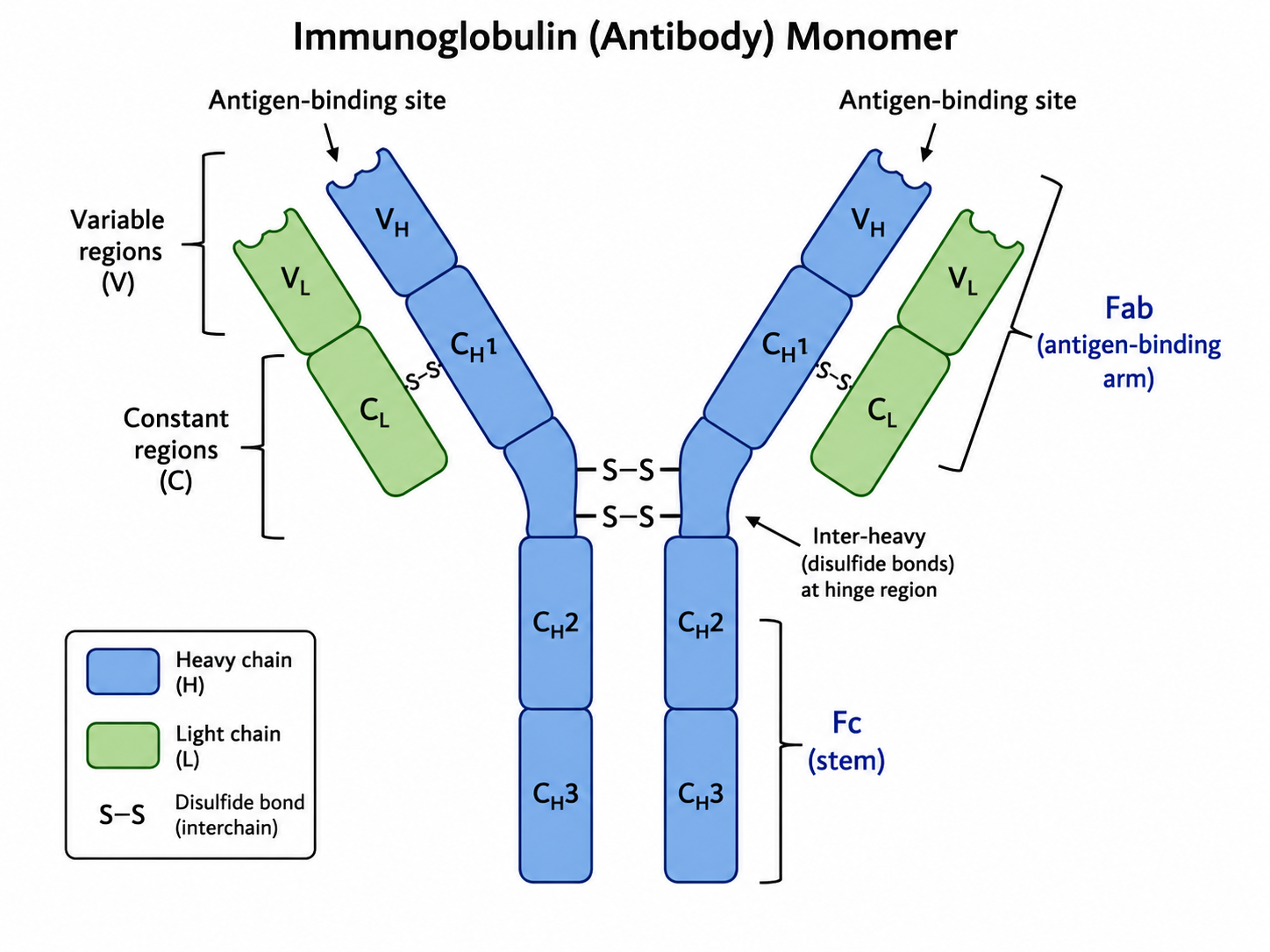
Must knowAn antibody is Y-shaped: two identical heavy chains + two identical light chains, held by disulfide bonds.
Each chain has a variable (V) region (amino terminus) and a constant (C) region (carboxyl terminus).
- The variable regions of one heavy + one light chain form the antigen-binding site (two per antibody — bivalent); the contact region is the complementarity-determining region (CDR).
- The constant heavy-chain region forms the Fc region — the "tail" that determines antibody class and effector function.
Functionally: Fab region (the two "arms" — variable regions and antigen-binding sites) and Fc region (the "stem" — effector functions).
Antibody Classes (Isotypes)
Must knowThe two high-yield classes:
- IgG — most abundant in serum; monomer; main player in opsonization and complement activation; only class that crosses the placenta.
- IgM — first antibody produced in a primary response; pentamer, making it especially effective at complement activation.
IgA (secretory/mucosal), IgE (mast cells, allergy/antiparasitic), IgD (naïve B-cell surface receptor).
Quick check: A mother who recovered from chickenpox years ago has a newborn. What antibody class protects the newborn in the first weeks of life, and how did it get there?
Answer: IgG — the only class that crosses the placenta (Fc receptor–mediated transcytosis), transported to the fetus during the third trimester, giving passive immunity that wanes over the first 3–6 months.
Antigen Presentation
Must knowThis concept links innate to adaptive immunity and is tightly tied to MHC I vs. II. When a pathogen is destroyed inside an APC, its peptide fragments are loaded onto MHC and displayed for T cells to inspect.
The key rule:
MHC class I presents endogenous peptides (made INSIDE the cell) → recognized by CD8+ cytotoxic T cells
MHC class II presents exogenous peptides (taken up from OUTSIDE) → recognized by CD4+ helper T cells
MHC Class I — "Inside Danger"
Must knowExpressed on all nucleated cells (not RBCs). Know the logic cytosolic proteins are degraded by the proteasome, transported into the ER by TAP, loaded onto MHC I, and displayed. Viral or cancer peptides appearing on MHC I flag the cell for CD8+ CTL killing — any nucleated cell can be infected, so all must be inspectable.
MHC Class II — "Outside Danger"
Must knowExpressed only on professional APCs (macrophages, dendritic cells, B cells). Exogenous proteins are taken up, degraded in the phagolysosome, loaded onto MHC II, and displayed to CD4+ T cells.
The Two-Signal Model of T Cell Activation
Know the logicNaïve T cells require two signals to activate:
- Signal 1 — TCR binds antigen on MHC (specificity).
- Signal 2 — costimulation: B7 (CD80/CD86) on the APC binds CD28 on the T cell.
A T cell that gets Signal 1 without Signal 2 becomes anergic (silenced) — a key mechanism of peripheral tolerance.
Cross-Presentation
OptionalDendritic cells can cross-present exogenous antigen on MHC class I, letting them activate CD8+ T cells against viruses or tumors even when not themselves infected.
Quick check: Mycobacterium tuberculosis survives inside macrophage phagosomes. Would Tb antigens be presented on MHC class I or class II? Which T cell is activated, and what is the outcome?
Answer: Phagosomal (exogenous) antigens are presented on MHC class II, activating CD4+ helper T cells. Th1 cells then secrete IFN-γ, which activates macrophages to better kill intracellular bacteria. (Some Tb peptides can also cross-present on MHC I, but the classical pathway is MHC II → CD4+.)
Major Histocompatibility Complex (MHC)
Must knowMHC molecules present peptide antigens to T cells. In humans they are encoded by the HLA (Human Leukocyte Antigen) gene complex on chromosome 6 — the most polymorphic genes in the genome. This extreme diversity means almost no two people share an HLA type, which is why organ transplant matching requires HLA typing.
| Feature | MHC Class I | MHC Class II |
|---|---|---|
| Expressed on | All nucleated cells | Professional APCs only |
| Presents | Endogenous (cytosolic) peptides | Exogenous (phagocytic) peptides |
| Recognized by | CD8+ T cells | CD4+ T cells |
| Pathway | Proteasome → TAP → ER → surface | Phagolysosome → endosome → surface |
In thymic selection: positive selection commits CD4 cells to bind self-MHC II and CD8 cells to self-MHC I; negative selection deletes T cells that bind self-peptide/self-MHC too strongly.
Quick check: Why are transplanted organs rejected, and which immune cells lead this rejection?
Answer: Donor MHC (HLA) is seen as foreign by recipient T cells. Donor cells present non-self MHC I to recipient CD8+ CTLs, which destroy the graft; donor APCs can also activate recipient CD4+ helper T cells. Both drive rejection — which is why T cell–targeting immunosuppressants (e.g., cyclosporine, which inhibits IL-2 production) are given post-transplant.
Clonal Selection
Must knowBefore any pathogen enters, you already have millions of naïve B and T cells, each with a unique, randomly generated receptor. This diversity arises from V(D)J recombination — random selection and joining of variable-region gene segments — generating millions of specificities from a limited gene set (concept only; the recombinase enzymology is out of scope).
Clonal selection is how the immune system activates the specific clone(s) matching an antigen:
- Antigen enters the body.
- It binds and activates only the lymphocytes whose receptor matches it.
- The selected lymphocyte proliferates — clonal expansion — into thousands of copies.
- These differentiate into effector cells (plasma cells, CTLs) and memory cells.
This explains why adaptive immunity is specific and improves on re-exposure.
Primary vs. Secondary Immune Response
Must know- Primary response (first exposure): ~7–14 day lag; predominantly IgM, lower titer and affinity.
- Secondary (anamnestic) response (re-exposure): rapid (1–3 days), high-magnitude, predominantly high-affinity IgG. This is the basis of vaccination.
Quick check: A patient gets their first flu vaccine in October and a booster in November. Compare the antibody response after each dose in speed, magnitude, and antibody class.
Answer: First dose (primary): slow (~7–14 days), low-titer, mostly IgM. Second dose (secondary/anamnestic): rapid (~1–3 days), much higher titer, mostly high-affinity class-switched IgG. The amplification reflects clonal expansion of memory B cells from the first dose.
Antigen-Antibody Recognition
Must knowAntibodies bind antigens via non-covalent interactions (hydrogen bonds, electrostatic, van der Waals, hydrophobic) — highly specific (CDR fits epitope) and reversible.
Effector Functions of Antibody Binding
Must knowAntibody binding triggers downstream effects:
- Neutralization — antibodies block pathogens or toxins from binding host receptors, rendering them inert.
- Opsonization — antibodies (especially IgG) coat a pathogen; phagocyte Fc receptors bind the Fc region, boosting phagocytosis ("handles" to grab).
- Complement activation — antigen-bound IgM/IgG activate complement → MAC lysis plus opsonin/chemoattractant fragments.
- ADCC (antibody-dependent cell-mediated cytotoxicity) — antibody-coated target cells are killed by NK cells binding the Fc region.
- Agglutination/Precipitation — bivalent/multivalent antibodies crosslink antigens into clumps that are more easily phagocytosed.
Quick check: Which antibody is more effective at complement activation: a monomer or a pentamer? Explain.
Answer: The pentamer — IgM — is far more effective per molecule because its multiple Fc regions in close proximity allow C1q (first component of the classical pathway) to bind. A single IgM can activate complement; IgG requires several molecules bound nearby.
Recognition of Self vs. Nonself; Autoimmune Diseases
Must knowNot attacking your own body is immunological tolerance, operating at two levels.
Central Tolerance
Must knowIn primary lymphoid organs during development: in the thymus, autoreactive T cells are deleted by negative selection; in the bone marrow, autoreactive B cells undergo receptor editing or clonal deletion.
Peripheral Tolerance
Know the logicCatches self-reactive lymphocytes that escape central tolerance:
- Anergy — T cells that meet antigen without costimulation are silenced.
- Regulatory T cells (Tregs) — suppress other lymphocytes.
- Clonal ignorance — some self-antigens are sequestered in immune-privileged sites (eye, brain, testes).
Autoimmune Diseases
Know the logicWhen tolerance fails, the immune system attacks self-tissues.
| Disease | Target |
|---|---|
| Type 1 Diabetes | Pancreatic β cells (CD8+ CTLs) |
| Rheumatoid Arthritis | Synovial joints |
| Multiple Sclerosis | CNS myelin |
| Myasthenia Gravis | Acetylcholine receptors (NMJ) |
| Graves' / Hashimoto's | Thyroid (hyper- / hypothyroid) |
| Lupus (SLE) | Nuclear antigens (anti-nuclear antibodies) |
Molecular mimicry is one mechanism: a pathogen antigen resembles a self-antigen, so clearing the pathogen also targets self (e.g., rheumatic fever — anti-Streptococcal M protein cross-reacts with cardiac muscle). Passage-level certain HLA alleles raise risk for specific autoimmune diseases, linking MHC to self-reactivity.
Quick check: Myasthenia gravis involves autoantibodies against acetylcholine receptors at the NMJ. Would you expect too much or too little muscle contraction? What drug class might help?
Answer: Too little — antibodies block/internalize ACh receptors, so released ACh cannot bind effectively → weakness, worse with repetitive use. Acetylcholinesterase inhibitors (e.g., pyridostigmine) help by raising synaptic ACh to compete with the blocking antibodies.
Active vs. Passive Immunity
Must knowActive immunity — your own immune system mounts a response and forms memory. Natural (infection) or artificial (vaccination). Slow to develop, long-lasting.
Passive immunity — pre-formed antibodies from another source; no memory. Immediate but short-lived.
| Type | Example | Memory? |
|---|---|---|
| Natural active | Recovering from chickenpox | Yes |
| Artificial active | Vaccine | Yes |
| Natural passive | IgG across placenta; IgA in breast milk | No |
| Artificial passive | Antivenom, immune globulin, monoclonal antibodies | No |
Common Confusions & Tricks
1. CD4/MHC II vs. CD8/MHC I — never mix these up.
The numbers multiply to 8: "CD 4 × MHC II = 8; CD 8 × MHC I = 8." Also: Exogenous → MHC II → CD4+ (helper); Endogenous → MHC I → CD8+ (cytotoxic). CD8 cells kill internally compromised cells, so they inspect what's made inside (endogenous = MHC I).
2. B cells recognize native antigen; T cells only recognize MHC-presented peptides.
B cells bind whole, free, unprocessed antigen via BCR. T cells cannot — they see only short peptides on MHC.
3. NK cells vs. CTLs — both kill cells, very differently.
NK cells are innate, need no antigen-specific activation, and kill cells that LACK MHC I. CTLs are adaptive, require MHC I + foreign peptide. Complementary: viruses that downregulate MHC I to dodge CTLs become vulnerable to NK cells.
4. IgM vs. IgG — first vs. best.
IgM is made FIRST (primary response, pre–class switch) and is a pentamer (great for complement). IgG comes LATER — crosses placenta, longer half-life, highest affinity, most abundant. Early infection → IgM; long-term/secondary → IgG.
5. Opsonization is NOT neutralization.
Neutralization: antibody blocks pathogen function (e.g., viral entry). Opsonization: antibody coats the pathogen so phagocytes eat it more easily.
6. Plasma cells have NO surface BCR — they only secrete antibody.
If a passage says "no surface Ig but secretes large amounts of antibody," that's a plasma cell.
7. Antigen vs. epitope.
An antigen is the whole molecule; an epitope is the specific region bound. One antigen has many epitopes → a polyclonal response uses multiple clones targeting different epitopes.
8. Primary lymphoid organs (bone marrow, thymus) ≠ secondary (lymph nodes, spleen, tonsils).
Lymphocytes DEVELOP in primary organs; responses are EXECUTED in secondary organs. T cells mature in the thymus, NOT lymph nodes.
9. Vaccination generates ACTIVE immunity — weakened/killed pathogen or antigen, not preformed antibodies. Antivenom and immune globulin are passive.
10. Autoimmune disease ≠ immunodeficiency.
Autoimmunity = overactive, misdirected response; immunodeficiency = weakened/absent. Opposites. HIV → immunodeficiency; lupus → autoimmunity.
Key Takeaways
Innate vs. Adaptive
- Innate: fast, non-specific, no memory; PRRs (TLRs) detect PAMPs
- Adaptive: slow initially, highly specific, generates memory; lymphocytes with rearranged receptors
Key Cell Types
- Neutrophils: first responders, phagocytosis + oxidative burst; elevated in bacterial infection
- Macrophages: phagocytosis, antigen presentation (MHC II), cytokines
- Dendritic cells: best APCs; bridge innate to adaptive
- NK cells: innate; kill cells lacking MHC I; perforin/granzyme
- Mast cells/basophils: IgE-mediated histamine release; allergy
- CD4+ helper T cells: MHC II; orchestrate via cytokines; HIV target; includes Tregs
- CD8+ cytotoxic T cells: MHC I; kill infected/cancerous cells; perforin/granzyme/Fas-FasL
- B cells/Plasma cells: B cells recognize native antigen; plasma cells secrete antibody; class switching → IgG, IgA, IgE from IgM
MHC — Critical Rules
- MHC I: all nucleated cells; endogenous peptides; CD8+ T cells
- MHC II: professional APCs only; exogenous peptides; CD4+ T cells
- HLA = human MHC; basis for transplant matching and autoimmune susceptibility
Antibody Classes
- IgG: most abundant, crosses placenta, opsonization + complement
- IgM: pentamer, first produced, best early complement activator
- (Recognition: IgA = mucosal; IgE = allergy/antiparasitic; IgD = naïve B-cell receptor)
Antibody Structure
- 2 heavy + 2 light chains; disulfide bonds; Y-shaped
- Fab: antigen binding (variable regions, CDRs); Fc: effector functions
Key Processes
- Clonal selection: antigen selects pre-existing clones; clonal expansion follows
- Primary response: slow, IgM; Secondary: fast, high-titer IgG — basis of vaccination
- Positive selection (thymus cortex): keep T cells that bind self-MHC
- Negative selection (thymus medulla): delete T cells that bind self-peptide/MHC too strongly (central tolerance)
- Antigen presentation: pathogen peptides displayed on MHC for T cell inspection
Lymphoid Organs
- Primary (development): bone marrow (B cells, hematopoiesis), thymus (T cells)
- Secondary (responses): lymph nodes (filter lymph), spleen (filter blood), tonsils/MALT
Active vs. Passive Immunity
- Active (infection or vaccination): slow onset, long-lasting, memory
- Passive (maternal IgG, antivenom, immune globulin): immediate, short-lived, no memory