The respiratory system handles gas exchange, helps regulate blood pH (by controlling how much is retained), and defends against inhaled particles and pathogens. The MCAT tests it across disciplines — from the physics of breathing to the chemistry of transport — and it integrates tightly with the cardiovascular, renal, and nervous systems.
Priority labels: Must know = cold; Know the logic = mechanism not names; Passage-level = recognize, don't memorize; Optional = skippable.
General Function and Overview
Must knowThree functions to keep in mind:
- Gas exchange — into blood, out
- pH regulation — adjusting how much is retained or expelled
- Protection — filtering, warming, and humidifying incoming air
These integrate with pulmonary circulation, renal pH control, and the respiratory drive. Passages often require reasoning across these systems at once.
Quick check: If a patient hyperventilates, what happens to blood and pH?
Answer: Blood falls (blown off), less carbonic acid forms, so pH rises (respiratory alkalosis). This links breathing mechanics to acid-base balance.
Structure of the Lungs and Airways
Anatomical Organization
Must knowAir travels: nasal cavity (or mouth) → pharynx → larynx (vocal cords) → trachea → splits into left/right primary bronchi → progressively branching bronchi → bronchioles → terminal bronchioles → alveoli. This branching bronchial tree increases total cross-sectional area distally, lowering air velocity — ideal for slow diffusion at the alveoli.
Know the logicThe conducting zone (down to terminal bronchioles) is anatomical dead space (~150 mL): it moves air but doesn't exchange gas. Physiological dead space adds alveoli that are ventilated but not perfused (e.g., pulmonary embolism). Because dead space is subtracted each breath, rapid shallow breathing (low tidal volume) wastes a large fraction of every breath — it ventilates airways but moves little fresh air to alveoli. (Formula: alveolar ventilation .)
Structure of the Alveoli
Must knowThe alveoli are tiny sacs at the end of the tree, built for gas exchange:
- Type I pneumocytes — thin squamous cells, ~95% of surface area; minimize diffusion distance.
- Type II pneumocytes — produce surfactant; also act as stem cells for Type I cells.
- Alveolar macrophages — phagocytose particles/pathogens reaching the alveoli.
- A dense capillary network wraps each alveolus; the blood–gas barrier is only ~0.5 μm thick.
The lungs sit in the thoracic cavity, each wrapped in a double-layered pleura (visceral on the lung, parietal on the chest wall) enclosing a thin-fluid pleural space. The resulting surface tension keeps the lung expanded against the chest wall — central to breathing mechanics.
Quick check: Why is air in the pleural space (pneumothorax) dangerous?
Answer: It destroys the pressure differential holding the lung to the chest wall. Lung elastic recoil now has no opposing outward force, so the lung collapses and gas exchange fails.
Breathing Mechanics
The Physics: Boyle's Law
Must knowBreathing is mechanical, governed by Boyle's Law (, or ). Expanding the thoracic cavity raises lung volume, dropping alveolar pressure below atmospheric → air flows in. Decreasing thoracic volume raises pressure above atmospheric → air flows out.
Inspiration vs. Expiration
Must knowInspiration is always active. The diaphragm contracts and flattens (increasing vertical dimension) and the external intercostals lift the ribs up and out — together increasing thoracic volume, dropping alveolar pressure, drawing air in. Forced inspiration recruits accessory muscles (sternocleidomastoid, scalenes).
Quiet expiration is passive — muscles relax and elastic recoil drives air out. Forced expiration is active, using internal intercostals and abdominal muscles.
The Key Pressures
Must knowIntrapleural pressure is always subatmospheric at rest. Transpulmonary pressure is what keeps the lungs inflated; lung collapse occurs when this gradient disappears (pneumothorax). (Atmospheric ≈ 760 mmHg; alveolar ≈ atmospheric at rest, dropping slightly on inhalation; intrapleural ≈ −4 cmH₂O gauge.)
Quick check: A paralytic drug stops all respiratory muscle activity. What happens to lung volume?
Answer: The lungs settle at FRC — the resting volume where inward lung recoil balances outward chest-wall recoil.
Resiliency, Compliance, and Surface Tension
Lung Compliance
Know the logicCompliance () is how easily the lung stretches. High compliance = expands easily; low (stiff) = needs more pressure.
- Emphysema: destroyed elastin → too compliant, traps air.
- Fibrosis / RDS: stiff, low-compliance lungs that are hard to inflate.
Surface Tension and Surfactant
Know the logicThe water film lining each alveolus creates surface tension that tends to collapse the sphere. The Law of Laplace gives the pressure needed to keep it open:
Smaller alveoli (small ) need greater pressure to stay open, so they tend to collapse into larger ones. Surfactant (a phospholipid from Type II pneumocytes) reduces surface tension — and does so more in smaller alveoli, equalizing pressures and preventing collapse. Without it, breathing would require enormous effort.
Passage-levelNeonatal RDS occurs in premature infants (~before 32 weeks) lacking surfactant; treated with exogenous surfactant and glucocorticoids.
Quick check: Two connected alveoli — one small, one large — both lack surfactant. What happens?
Answer: By Laplace's Law the smaller alveolus has higher internal pressure, so it pushes air into the larger one and collapses. Surfactant prevents this by lowering surface tension more in the smaller alveolus.
Protection Against Disease: Filtration Systems
Must knowThe nasal cavity is the first filter: nasal hairs (vibrissae) trap large particles and turbinates create turbulence that drives particles into mucus, while the mucosa warms and humidifies air.
The trachea and bronchi use the mucociliary escalator: goblet cells secrete mucus that traps particles, and cilia beat in a coordinated wave to propel it upward toward the pharynx to be swallowed or expelled. Smoking paralyzes and destroys cilia, disabling clearance (smoker's cough, more infections). Particles reaching the alveoli are cleared by alveolar macrophages.
Quick check: In Kartagener's syndrome cilia are immotile (dynein arm defect). What respiratory symptom would you expect?
Answer: Chronic, recurrent respiratory infections — the mucociliary escalator can't clear mucus and trapped pathogens.
Thermoregulation: Nasal/Tracheal Capillary Beds, Evaporation, and Panting
Know the logicThe richly vascularized nasal mucosa (with countercurrent exchange in the turbinates) warms inhaled air toward body temperature and humidifies it; on exhalation, cooler air recovers heat and water from the mucosa, conserving both. Tracheal capillary beds continue this, so air reaches the alveoli warmed and fully saturated. (Because air is humidified and is continuously absorbed, alveolar ~100 mmHg is below dry atmospheric.)
Passage-levelPanting: in animals like dogs, rapid shallow breathing increases evaporation from the oral mucosa to dissipate heat. It stays shallow (ventilating dead space) to avoid blowing off .
Quick check: Why does air at altitude have a lower effective even though stays 21%?
Answer: Partial pressure = fraction × total pressure. Lower atmospheric pressure at altitude lowers inspired and alveolar , shrinking the diffusion gradient into blood — causing hypoxia.
Alveolar Gas Exchange
Diffusion: Fick's Law
Must knowGas exchange is entirely passive diffusion. Fick's Law:
where = surface area, = diffusion coefficient (∝ solubility), = partial-pressure gradient, = membrane thickness. The alveolar–capillary interface optimizes all four: huge surface area (~70 m²), large gradients, and an ultra-thin (~0.5 μm) membrane.
Partial Pressures
Must knowKnow the pattern, not every number. Gases move down partial-pressure gradients:
- Lungs: from alveolus (~100) → blood (~40); from blood (~46) → alveolus (~40).
- Tissues: from blood (~100) → tissue (~40); from tissue (~46) → blood (~40).
Alveolar (~100) is below atmospheric (~160) because alveolar air is diluted by water vapor/residual gas and is continuously absorbed.
diffuses ~20× more readily than (far more soluble), so diffusion-impairing disease (fibrosis, edema) causes hypoxia before hypercapnia.
Henry's Law
Must knowHenry's Law: dissolved gas is proportional to its partial pressure (). Key applications:
- Higher partial pressure → more dissolved gas (basis of hyperbaric ; divers dissolve more at depth).
- Decompression sickness ("the bends"): ascending too fast lets dissolved bubble out of solution.
- decreases with temperature — warm liquids hold less dissolved gas.
Quick check: A patient in a hyperbaric chamber at 3 atm breathes 100% . What happens to dissolved plasma ?
Answer: By Henry's Law, rises far above normal (~2280 vs ~100 mmHg), so dissolved plasma increases dramatically — supplying tissues independent of hemoglobin. This is the basis of hyperbaric therapy.
Oxygen Transport on Hemoglobin
Must knowOnly a tiny fraction of travels dissolved; most binds hemoglobin, a tetramer whose four subunits each bind one . Cooperative binding (one raises affinity of the others) makes the saturation-vs- curve sigmoidal, letting hemoglobin load fully in the lungs (high ) yet unload readily in tissues (low ).
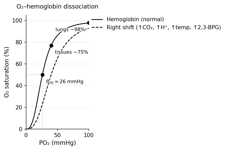
A rightward shift = lower affinity = favors unloading to tissues. Four factors shift it right — exactly the conditions of active tissue:
- ↑ · ↑ (↓ pH) · ↑ temperature · ↑ 2,3-BPG
The pH/ portion is the Bohr effect: in exercising tissue, acid and accumulate → right shift → more released where needed. In the lungs the reverse loads .
Quick check: During intense exercise a muscle's temperature and rise. What happens to delivery there?
Answer: Both shift the curve right (lower affinity), so hemoglobin unloads more at the same tissue — delivering more to the most active muscle (Bohr effect).
CO₂ Transport and pH Control
Three Forms of CO₂ Transport
Must knowtravels to the lungs three ways:
- Dissolved in plasma (~7–10%) — small, but this fraction sets and thus pH.
- Carbaminohemoglobin (~20%) — bound to hemoglobin's amino groups (not heme iron, unlike ).
- Bicarbonate () (~70%) — the major form.
Bicarbonate Buffer System
Must knowIn red blood cells, carbonic anhydrase catalyzes:
At tissues, high drives this right; exits the RBC in exchange for (chloride shift), and the is buffered by hemoglobin. At the lungs everything reverses and is exhaled. (The Haldane effect: deoxygenated hemoglobin accepts better.)
Henderson-Hasselbalch
Must know
The ratio sets pH; the numerator is metabolic, the denominator respiratory. Know the four disturbances:
- Respiratory acidosis: → pH (hypoventilation, COPD, opioids)
- Respiratory alkalosis: → pH (hyperventilation, altitude)
- Metabolic acidosis: → pH; lungs compensate by hyperventilating
- Metabolic alkalosis: → pH; lungs compensate by hypoventilating
Worked example: pH 7.52, 30, 24. The pH is high (alkalosis), is low (the driver), and is normal (kidneys haven't compensated) → acute primary respiratory alkalosis.
Regulation of Breathing by the Nervous System
Medullary Respiratory Centers
Know the logicBreathing is driven by a central pattern generator in the brainstem (medulla and pons).
In the medulla, the Dorsal Respiratory Group (DRG) sets the basic inspiratory rhythm (firing the diaphragm via the phrenic nerve), and the Ventral Respiratory Group (VRG) is recruited for forced breathing. In the pons, the pneumotaxic center limits inspiration duration (switches it off) while the apneustic center prolongs it. (Pre-Bötzinger complex = the core rhythm generator.)
CO₂ Sensitivity: The Primary Driver
Must knowThe primary stimulus for breathing is (really pH), not .
Central chemoreceptors (ventral medulla) don't detect blood directly — they detect CSF pH. (but not or ) crosses the blood-brain barrier, forms carbonic acid, lowers CSF pH, and stimulates ventilation:
Peripheral chemoreceptors in the carotid bodies (via CN IX) and aortic bodies (via CN X) respond to (only once it drops below ~60 mmHg), , and pH directly.
Why , not : isn't sensed until it falls below ~60 mmHg (hemoglobin is still ~90% saturated there), whereas a small rise drives a powerful ventilatory response.
Passage-levelCOPD "hypoxic drive": chronic retainers desensitize their central chemoreceptors and come to rely on hypoxia (peripheral) to breathe, so high-flow can blunt their drive. (Clinically nuanced, but this is what the MCAT tests.)
OptionalOther inputs: Hering-Breuer reflex (stretch receptors prevent overinflation), irritant receptors (cough), and voluntary cortical override.
Quick check: A lesion destroys only the peripheral chemoreceptors. The patient breathes normally at rest, then ascends to altitude. What happens?
Answer: A blunted hypoxic ventilatory response — without -sensitive peripheral chemoreceptors, they can't ramp up ventilation in response to the low of altitude.
Ventilation-Perfusion Matching (V/Q Ratio)
Must knowEfficient exchange needs alveoli both ventilated and perfused. Normal V/Q ≈ 0.8, varying by gravity: the apex has ventilation > perfusion (high V/Q), the base has perfusion > ventilation (low V/Q).
V/Q mismatch is the most common cause of hypoxia in lung disease:
- High V/Q (dead space): ventilated but not perfused (e.g., pulmonary embolism).
- Low V/Q (shunt): perfused but not ventilated (e.g., pneumonia, collapse).
Hypoxic pulmonary vasoconstriction: unlike systemic arterioles (which dilate when is low), pulmonary arterioles constrict when local alveolar falls, redirecting blood toward better-ventilated regions. (Chronic global hypoxia at altitude makes this widespread, raising pulmonary pressure and straining the right heart.)
Quick check: A pulmonary embolism causes which V/Q mismatch, and what happens to blood ?
Answer: High V/Q (dead space) — affected alveoli get no blood. The patient hyperventilates unaffected regions, blowing off enough (which diffuses readily) to keep normal or low, while stays low.
Common Confusions & Tricks
1. Inspiration decreases pressure; it does NOT actively "suck in" air. The diaphragm increases thoracic volume → alveolar pressure drops below atmospheric → air flows in down the gradient.
2. Surfactant prevents collapse, not expansion. It lowers surface tension so less pressure is needed to keep alveoli open. Without it, alveoli collapse on exhalation — why the first breath in RDS is hardest.
3. is the primary driver of breathing; is not. You breathe mainly to control /pH, not to get oxygen.
4. Central chemoreceptors detect CSF pH, not blood pH or directly. crosses the BBB → lowers CSF pH. Metabolic acidosis (blood , not ) is sensed more by peripheral chemoreceptors, since doesn't cross the BBB readily.
5. Henry's Law: decreases with temperature (opposite of most solubility). Warm soda goes flat faster.
6. Laplace's Law — smaller alveoli need higher pressure to stay inflated (not lower), which is why surfactant is critical.
7. The "dissolved" fraction is small (~7–10%) but sets and pH. Most travels as , but dissolved drives the acid-base chemistry.
8. Gases cross the alveolar membrane by diffusion ONLY — no transporters or pumps. Implied active transport is a trap.
Key Takeaways
Structure and Function
- Airways branch trachea → bronchi → bronchioles → alveoli; surface area ~70 m²
- Type I pneumocytes: gas exchange; Type II: surfactant + stem cells
- Pleural space maintains subatmospheric pressure that keeps lungs expanded
Breathing Mechanics
- Inspiration = active (diaphragm + external intercostals → ↑volume → ↓pressure → air in)
- Quiet expiration = passive (recoil); forced expiration = active (internal intercostals + abdominals)
- With no muscle activity, lungs settle at FRC (lung and chest-wall recoil balance)
Surface Tension and Surfactant
- Laplace's Law — smaller alveoli need more pressure to stay open
- Surfactant (Type II pneumocytes) reduces surface tension, stabilizes alveoli; absent → neonatal RDS
Gas Exchange
- Passive diffusion (Fick's Law): rate ∝ area × gradient / thickness
- Alveolar ≈ 100, venous ≈ 40 → into blood; ~20× more diffusible than
- Henry's Law ; decreases with temperature
- Most on hemoglobin; sigmoidal curve shifts right (unloads more) with ↑, ↑/↓pH, ↑temp, ↑2,3-BPG (Bohr effect)
CO₂ Transport and pH
- as: dissolved (~7%), carbaminohemoglobin (~20%), bicarbonate (~70%)
- Carbonic anhydrase: ; chloride shift moves out for
- Henderson-Hasselbalch: ; normal 7.40
Nervous Regulation
- Medulla: DRG (basic rhythm), VRG (forced); pons: pneumotaxic (limits) / apneustic (prolongs) inspiration
- Central chemoreceptors (medulla): detect ↓CSF pH from crossing BBB → primary drive
- Peripheral chemoreceptors (carotid/aortic): detect ↓, ↑, ↓pH → secondary; CN IX / CN X
- COPD retainers depend on hypoxic drive → use supplemental cautiously
Protection
- Nasal hairs → turbinates/mucus → mucociliary escalator → alveolar macrophages
- Cilia beat upward; destroyed by smoking → more infections
- Panting: shallow breathing ventilating dead space for evaporative cooling