Muscle cells are the body's force generators. Everything from pumping blood to lifting a textbook relies on the same trick: converting chemical energy (ATP) into mechanical movement by sliding protein filaments past each other. Keep that central idea as your anchor — the molecular details all serve that one purpose.
Priority labels: Must know = cold; Know the logic = mechanism not names; Passage-level = recognize, don't memorize; Optional = skippable.
Types of Muscle Tissue: The Big Picture
Must knowYou need a clear map of the three muscle types. The MCAT mixes-and-matches their features in passages, so know each one's structure, control, and function.
Skeletal (striated) muscle is voluntarily controlled (somatic motor neurons at the neuromuscular junction). It shows striations from the precise alignment of protein filaments. Each cell — a muscle fiber — is huge, runs much of the muscle's length, and has multiple peripheral nuclei (it's a syncytium formed by fusion of precursor myoblasts). Muscle-specific terms: cytoplasm = sarcoplasm, membrane = sarcolemma, specialized smooth ER = sarcoplasmic reticulum (SR), the intracellular Ca²⁺ store.
Cardiac muscle is found only in the heart and is also striated, but cells (cardiomyocytes) are smaller, branched, and have a single central nucleus. They join end-to-end at intercalated discs, which contain gap junctions (electrical coupling → coordinated contraction) and desmosomes (mechanical strength). Cardiac muscle is involuntary and autorhythmic (the SA node sets pace, autonomics modulate), highly fatigue-resistant, and mitochondria-rich.
Smooth muscle lines hollow organs and vessels (GI tract, blood vessels, uterus, bladder, bronchioles). It is non-striated (contractile proteins attach to dense bodies, not regular sarcomeres), with spindle-shaped cells and a single central nucleus. Control is involuntary (autonomic, hormones, paracrine). It contracts slowly but sustains tone for long periods without fatiguing. Key distinction the MCAT loves: smooth muscle does not use troponin — it uses calmodulin and myosin light-chain kinase (MLCK).
| Feature | Skeletal | Cardiac | Smooth |
|---|---|---|---|
| Striations | Yes | Yes | No |
| Nuclei per cell | Multiple, peripheral | 1–2, central | 1, central |
| Control | Voluntary | Involuntary | Involuntary |
| Intercalated discs | No | Yes | No |
| Fatigue resistance | Variable | Very high | Very high |
| Ca²⁺ regulation | Troponin/tropomyosin | Troponin/tropomyosin | Calmodulin/MLCK |
| Location | Attached to skeleton | Heart | Hollow organs, vessels |
Quick check: A biopsy shows cells with striations, branching, and intercalated discs with a single central nucleus. What muscle type is this, and where is it found?
Answer: Cardiac muscle, found only in the heart.
Red vs. White Muscle Fibers and the Role of Mitochondria
Must knowSkeletal fibers come in two flavors the MCAT cares about:
- Type I (slow-twitch, red): built for endurance. Rich in myoglobin (the O₂-binding protein that gives the red color) and mitochondria, so they regenerate ATP by oxidative phosphorylation → fatigue-resistant. Postural muscles, marathoners' legs.
- Type II (fast-twitch, white): built for speed/power. Fewer mitochondria, less myoglobin, depend more on glycolysis → fast but fatigue quickly. Sprinting, explosive lifting.
Abundant mitochondria are the primary ATP source for sustained contraction because oxidative phosphorylation yields far more ATP per glucose (~30–32) than anaerobic glycolysis (~2 net). Myoglobin acts as a local O₂ reservoir, binding O₂ with high affinity and releasing it when demand rises.
Quick check: A long-distance cyclist's leg muscles appear dark red on dissection. What structural feature accounts for this color, and what does it tell you about the metabolic strategy of these fibers?
Answer: High myoglobin content (and abundant mitochondria) gives red muscle its color. The fibers rely on aerobic/oxidative metabolism for sustained, fatigue-resistant ATP production.
Organization of Contractile Elements: Actin, Myosin, and the Cross-Bridge Cycle
Must knowHere is the heart of muscle physiology. Build the image from the inside out: each muscle fiber is filled with myofibrils (longitudinal bundles running the cell's length), each of which is a chain of repeating contractile units called sarcomeres. Myofibrils sit beside the SR (Ca²⁺) and mitochondria (ATP).
Thin filaments are built on F-actin — a double helix of polymerized G-actin monomers, each carrying a myosin-binding site (blocked at rest; see calcium regulation).
Thick filaments are bundles of myosin molecules. Each myosin has intertwined tails forming the backbone and globular myosin heads that project outward. The heads are the business end: both an ATPase and an actin-binding domain.
The Cross-Bridge Cycle
Must knowThe cycle by which myosin "walks" along actin to shorten the sarcomere. Learn the sequence cold:
- Cocked state: myosin head has hydrolyzed ATP (ADP + Pᵢ still bound) and is in its high-energy "cocked" conformation.
- Cross-bridge formation: the head binds the exposed actin site, forming a cross-bridge.
- Power stroke: Pᵢ release drives the head to swing to its low-energy position, pulling the thin filament toward the sarcomere center. ADP is then released. This is the force-generating step.
- Rigor state: still tightly bound to actin, ADP and Pᵢ gone — the basis of rigor mortis (no ATP after death → myosin stays locked).
- Detachment: a new ATP binds the head, causing it to detach; hydrolysis re-cocks the head and the cycle repeats.
ATP's two roles here: (1) hydrolysis re-cocks the head (energy for the next power stroke); (2) new ATP binding allows detachment. Without ATP → rigor; without calcium, the cycle never starts.
The net result of many cross-bridges cycling is that thin filaments are pulled inward — the sarcomere shortens.
Quick check: A patient dies and their muscles become rigid within hours. What molecular event explains this rigidity?
Answer: Without ATP (no respiration after death), myosin heads cannot detach from actin after the power stroke (detachment requires new ATP binding). Cross-bridges remain locked — rigor mortis.
Sarcomere Structure: Bands, Lines, and Zones
Must knowThe sarcomere is the repeating contractile unit, bounded at each end by a Z-line (Z-disc). Knowing its anatomy lets you predict how each landmark changes during contraction — a favorite MCAT question.
| Structure | What it is | During contraction |
|---|---|---|
| Z-line | Anchors thin filaments; sarcomere boundary | Move closer together |
| M-line | Center; cross-links thick filament tails | No change in position |
| A-band | Full length of thick (myosin) filaments; dark | Stays the same |
| I-band | Thin filaments only; light | Shortens |
| H-zone | Central A-band region, thick only (no thin overlap) | Shortens |
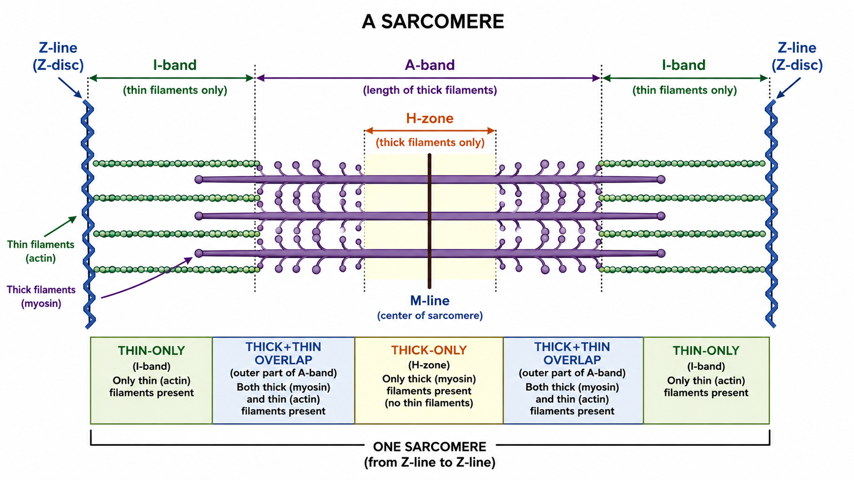
Sliding filament model: during contraction filament lengths do not change — thin filaments slide inward, overlapping more with thick filaments. So the I-band and H-zone shorten, the A-band stays constant (it equals thick-filament length), and Z-lines move closer.
Mnemonic: "I H8 contraction" — the I-band and H-zone shorten; the A-band is Always the same.
Quick check: A student claims that during maximum contraction the A-band shortens because the myosin filaments are compressed. Is this correct?
Answer: No. The A-band reflects myosin filament length, which doesn't change. The I-band and H-zone shorten (possibly to near zero); the A-band stays constant.
Troponin, Tropomyosin, and the Molecular Switch
Must knowActin's myosin-binding sites are blocked at rest by the troponin–tropomyosin complex; calcium is the key that unlocks them.
Tropomyosin is a long, rod-shaped protein lying in the F-actin grooves. At rest it sterically covers the myosin-binding sites — no cross-bridges form.
Troponin is a three-subunit complex:
- TnT binds tropomyosin (anchors the complex).
- TnI is the inhibitory subunit (holds tropomyosin in the blocking position).
- TnC is the calcium-binding subunit (Ca²⁺ binds here to trigger contraction; structurally similar to calmodulin).
Memory hook: Tropomyosin, Inhibitory, Calcium → TIC.
Quick check: Cardiac troponins (cTnI, cTnT) are released into the blood when cardiomyocytes are damaged. Why are these biomarkers so specific to cardiac damage, and why does the MCAT care?
Answer: Cardiac muscle expresses unique TnI/TnT isoforms not found elsewhere. Elevated serum troponin is the gold-standard biomarker for myocardial infarction — a classic MCAT link from biochemistry to clinical medicine.
Calcium Regulation of Contraction
Must knowCalcium is the master on/off switch for striated-muscle contraction. The whole sequence from nerve impulse to shortening sarcomere is excitation-contraction coupling.
The sequence:
- Neural signal: an action potential reaches the NMJ; acetylcholine is released, binds nicotinic receptors on the sarcolemma, and triggers a muscle action potential.
- T-tubules carry it inward: the action potential spreads along the sarcolemma and into transverse tubules (T-tubules) — invaginations that reach the fiber's core, so every myofibril is signaled at once.
- Calcium release: T-tubule depolarization opens SR Ca²⁺ release (via DHPR–ryanodine receptor coupling), flooding the sarcoplasm with Ca²⁺. Know the logic in cardiac muscle, a small Ca²⁺ entry triggers further SR release — calcium-induced calcium release.
- Ca²⁺ binds TnC.
- Tropomyosin shifts, uncovering actin's myosin-binding sites.
- Cross-bridge cycle begins → sarcomere shortens.
- Relaxation: when signaling stops, SERCA (an SR Ca²⁺-ATPase) actively pumps Ca²⁺ back into the SR. As cytosolic Ca²⁺ falls, tropomyosin re-covers the sites and the muscle relaxes.
Key connection: both contraction (power stroke) and relaxation (SERCA pumping) require ATP. No ATP → can't cross-bridge cycle and can't relax → prolonged contraction/cramping, ultimately rigor.
Smooth muscle — the contrast: smooth muscle lacks troponin. Rising Ca²⁺ binds calmodulin; the Ca²⁺–calmodulin complex activates MLCK, which phosphorylates myosin light chains to enable contraction. (Phosphatase reverses it — details are out of scope.)
Quick check: A drug blocks SERCA in a skeletal muscle fiber. Predict the consequence for muscle function.
Answer: Ca²⁺ can't return to the SR, so cytosolic Ca²⁺ stays high, TnC stays occupied, and binding sites stay exposed — the muscle remains contracted (cannot relax) until Ca²⁺ is removed by other means or ATP is depleted.
Common Confusions & Tricks
1. A-band stays the SAME; I-band and H-zone shorten. Remember A-band = Always the same (it equals myosin length). The I-band and H-zone are the variable regions that shrink.
2. Troponin is striated-muscle ONLY; smooth muscle uses calmodulin/MLCK. If a passage asks about Ca²⁺ regulation in smooth muscle, troponin is the wrong answer.
3. ATP causes detachment, not the power stroke directly. Hydrolysis cocks the head; the power stroke is driven by conformational change on Pᵢ release after actin binding. New ATP binding causes detachment.
4. Rigor mortis ≠ lack of calcium. Rigor = no ATP to release myosin from actin. Don't confuse it with failure to initiate contraction (calcium-dependent).
5. Myoglobin ≠ hemoglobin. Both are heme O₂-binders, but myoglobin is a monomer with a hyperbolic curve (storage in muscle); hemoglobin is a tetramer with a sigmoidal curve (cooperativity). Myoglobin's higher O₂ affinity lets it accept O₂ from hemoglobin.
6. Cardiac troponin as a biomarker. Cardiac-specific cTnI/cTnT isoforms are released when cardiomyocytes die — the basis for measuring them in suspected MI.
7. "Thin = Actin, Thick = Myosin" — never mix these up. Thin = actin (+ troponin + tropomyosin); thick = myosin. Confusing them breaks all band/zone interpretation.
8. Gap junctions in cardiac muscle. Intercalated discs contain gap junctions (electrical coupling) AND desmosomes (mechanical coupling).
Key Takeaways
- Three muscle types: skeletal (striated, multinucleate, voluntary), cardiac (striated, branched, intercalated discs with gap junctions, involuntary), smooth (non-striated, spindle-shaped, involuntary).
- Red (Type I) fibers = myoglobin + mitochondria → aerobic, fatigue-resistant; white (Type II) = glycolytic, fast, fatigue quickly.
- Thin filaments = actin + tropomyosin + troponin (TnT, TnI, TnC); thick filaments = myosin (heads = ATPase + actin-binding).
- Sarcomere = Z-line to Z-line. A-band constant; I-band and H-zone shorten; M-line anchors thick filaments at center.
- Sliding filament model: filament lengths don't change; thin filaments slide toward the M-line, shortening the sarcomere.
- Cross-bridge cycle: cocked head (ADP+Pᵢ) binds actin → Pᵢ release → power stroke → ADP release (rigor) → new ATP binds → detachment → re-cock. Repeat.
- ATP roles: (1) re-cock the head, (2) detach myosin from actin, (3) power SERCA for relaxation.
- Rigor mortis = no ATP → myosin can't detach.
- Striated Ca²⁺ regulation: SR Ca²⁺ release → Ca²⁺ binds TnC → tropomyosin uncovers sites → contraction; SERCA pumps Ca²⁺ back → relaxation.
- Smooth muscle: no troponin; Ca²⁺ → calmodulin → MLCK → contraction.
- Cardiac biomarker: cTnI/cTnT released during MI.