Spectroscopy lets you deduce a molecule's structure by measuring which photon energies it absorbs. The MCAT tests three windows—infrared (IR), ultraviolet-visible (UV-Vis), and nuclear magnetic resonance (NMR)—each probing a different structural feature.
Priority labels: Must know = cold; Know the logic = mechanism not names; Passage-level = recognize, don't memorize; Optional = skippable.
The Big Picture: Why Different Energies Probe Different Features
Must knowA molecule's bonds vibrate, its electrons occupy orbitals, and its nuclei act like tiny magnets—each motion takes a different energy to perturb. When a photon's energy exactly matches the gap between two states, the molecule absorbs it; everything else passes through. That selectivity is what makes spectroscopy structurally informative.
The energy ordering: NMR (radiowave, nuclear spin) < IR (bond vibration) < UV-Vis (electronic transition). Photon energy is
so higher frequency (shorter ) = more energetic photon. That's why electronic transitions (UV-Vis) need shorter wavelengths than vibrations (IR), which need shorter wavelengths than nuclear-spin flips (NMR).
Infrared Spectroscopy
Intramolecular Vibrations and Rotations
Must knowModel a bond as two masses on a spring. Its vibration frequency depends on spring stiffness (bond strength/order) and atomic masses (Hooke's Law):
where is the wavenumber (cm), the force constant (bond strength), and the reduced mass.
Two takeaways:
- Stronger/higher-order bonds vibrate at higher wavenumber: C≡C > C=C > C–C.
- O–H and N–H absorb above C–H mainly because they have a higher force constant (stronger, more polar)—not reduced mass, since for any X–H bond amu (dominated by H). The mass-driven case is C–H vs C–D: deuterium doubles , lowering the wavenumber (basis of isotopic labeling).
Selection rule: a vibration is IR-active only if it changes the dipole moment. The oscillating dipole couples to the photon's electric field. Symmetric stretches with no dipole change (, , the symmetric stretch of ) are IR-silent. Polar bonds (C=O, O–H, N–H) absorb strongly.
Vibration types: stretching (bond length changes; the most diagnostic peaks) and bending (bond angles change; lower energy, dominates the fingerprint region).
IR spectra plot transmittance (%) on y vs. wavenumber (cm) on x, decreasing left (~4000) to right (~400). A peak pointing down = absorption.
Quick check: If you replace ethanol's –OH with –OD, does the O–D stretch appear at higher or lower wavenumber than O–H? Answer: Lower. Deuterium doubles the reduced mass , and decreases.
Recognizing Common Characteristic Group Absorptions and the Fingerprint Region
Must knowThe spectrum has two zones.
Functional group region (4000–1500 cm): diagnostic stretches. Know these:
| Bond / Group | Region (cm) | Key Feature |
|---|---|---|
| O–H (alcohol) | 3200–3550 | Broad (H-bonding) |
| O–H (carboxylic acid) | 2500–3300 | Very broad, overlaps C–H |
| N–H (amine/amide) | 3300–3500 | 1° = two peaks, 2° = one |
| C–H (sp³ / sp²–sp) | <3000 / >3000 | Splits at 3000 cm |
| C≡N, C≡C | 2100–2260 | Sharp |
| C=O carbonyl | 1700 (1630–1815) | Strong, sharp; position = class |
| C=C alkene | ~1650 | Moderate |
Carbonyl position is the highest-yield IR pattern: the C=O wavenumber rises as C=O bond order rises. Electron donation into C=O (amide N lone pair) lowers it; electron withdrawal (Cl) raises it.
| Compound Class | C=O Wavenumber |
|---|---|
| Amide | ~1650 (lowest) |
| Carboxylic acid | ~1710 |
| Aldehyde / Ketone | ~1715–1745 |
| Ester | ~1735–1750 |
| Acid chloride / Anhydride | ~1800 (highest) |
Fingerprint region (400–1500 cm): complex bends and C–X stretches, unique per molecule. Don't interpret peak-by-peak; its use is comparison—identical fingerprints = same compound.
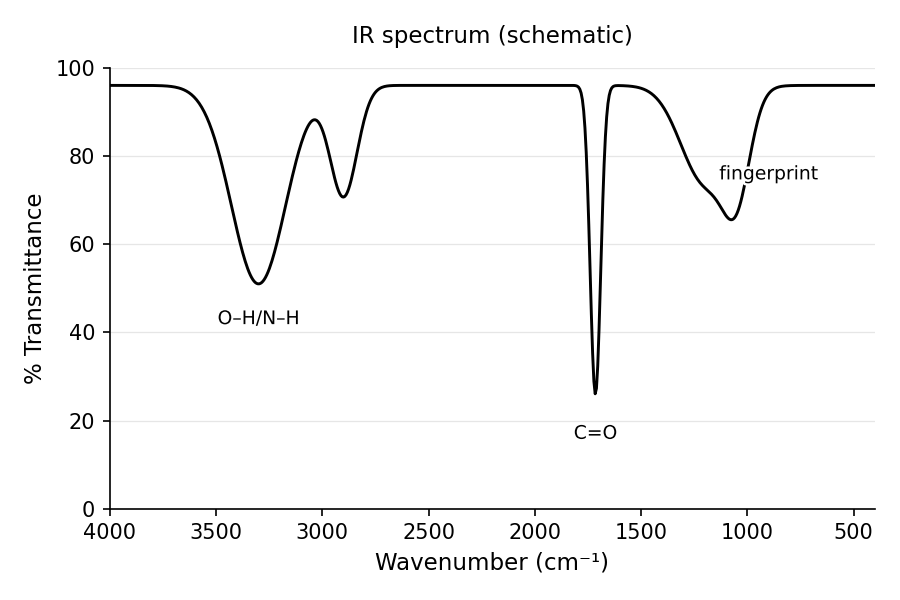
Quick check: An IR shows a very broad absorption spanning 2500–3300 cm and a carbonyl at 1710 cm. What group is present? Answer: Carboxylic acid—the broad H-bonded O–H plus a ~1710 C=O is the classic signature.
Ultraviolet-Visible (UV-Vis) Spectroscopy
-Electron and Nonbonding Electron Transitions
Must knowUV-Vis promotes valence electrons between molecular orbitals. Three relevant types:
- electrons (single bonds): very high energy (far UV, not relevant).
- electrons (double/triple bonds, aromatics): undergo .
- n electrons (lone pairs on O, N, S, halogens): undergo .
is allowed = intense band; is symmetry-forbidden = weak band at longer wavelength.
A chromophore is the part responsible for absorption (conjugated system or C=O). An auxochrome is a substituent (–OH, –NH) that shifts without absorbing strongly itself.
Quick check: A carbonyl shows a weak absorption at 310 nm. or ? Answer: —weak and at longer wavelength, the symmetry-forbidden lone-pair promotion.
Conjugated Systems and the Effect of Structural Changes
Must knowThe central idea: conjugation lowers the HOMO–LUMO gap, shifting absorption to longer wavelength. Each added conjugated double bond gives a bathochromic (red) shift; losing conjugation gives a hypsochromic (blue) shift. (Particle-in-a-box intuition: a longer "box" has closer-spaced levels—no quantitative formula needed.)
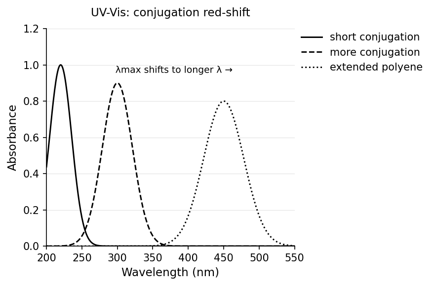
The qualitative trend (you do not need Woodward–Fieser increments): isolated C=C absorbs deep in UV (~180 nm); a conjugated diene at ~220–250 nm; extended polyenes reach into the visible; benzene ~250–280 nm.
Color: -carotene's 11 conjugated double bonds absorb blue-violet (~450–480 nm), so we see the complementary color, orange (why carrots are orange). For any colored molecule, you see what is not absorbed.
Passage-levelComplementary color pairs (recognize, don't memorize the full table): violet↔yellow, blue↔orange, green↔red, yellow↔violet, red↔blue-green.
Know the logicIndicators: pH indicators change conjugation with protonation state. Phenolphthalein is colorless in acid (lactone form, broken conjugation) and pink in base ( opens the lactone to a fully conjugated trianion). The structural change extends the system, shifting into the visible. General rule: extending conjugation red-shifts, disrupting it blue-shifts.
Beer-Lambert Law (know the relationship; quantitative work is Passage-level):
= absorbance, = molar absorptivity (L mol cm), = path length (cm), = concentration (mol L). Absorbance and transmittance:
Worked Example: A dye in a 1.00 cm cuvette gives , with L mol cm. Find .
Sanity check: , so ~75% absorbed—reasonable for a tinted solution.
NMR Spectroscopy
Protons in a Magnetic Field and Equivalent Protons
Must knowA H nucleus (spin 1/2) can align with (lower energy) or against a magnetic field; the gap is in the radiowave region and scales with field strength . RF energy matching the gap makes protons resonate (flip spin). The exact frequency depends on the local electronic environment: surrounding electrons partly shield each proton, so
More shielding → lower frequency → upfield (lower chemical shift ); less shielding (near electronegative atoms) → downfield (higher ).
is in ppm relative to TMS (). All organic protons appear downfield of TMS.
Canonical shift ranges:
| Proton Environment | (ppm) |
|---|---|
| Alkyl (CH, CH) | 0.9–1.5 |
| α to C=O | 2.0–2.7 |
| Next to N/O (–OCH) | 3.0–4.0 |
| Vinyl (C=C–H) | 4.5–6.5 |
| Aromatic (Ar–H) | 6.5–8.5 |
| Aldehyde (–CHO) | 9–10 |
| Carboxylic acid (–COOH) | 10–12 |
Aromatic protons are especially downfield because the benzene ring current deshields the H atoms.
Equivalent protons: protons interconvertible by molecular symmetry are chemically equivalent and give one signal (e.g., the 3 H of CHCl; the 6 H of benzene). The number of non-equivalent environments = number of signals.
Worked Example: How many H signals does ethyl acetate () show? Three environments—acetyl CH (~2.0), –OCH– (~4.1), ethyl CH (~1.2). Answer: 3 signals. The –OCH– is most downfield (bonded to O).
Quick check: How many H signals does para-xylene show? Answer: Two—the four aromatic H's are equivalent by symmetry (one signal ~7 ppm) and the two methyls are equivalent (~2.3 ppm).
Spin-Spin Splitting
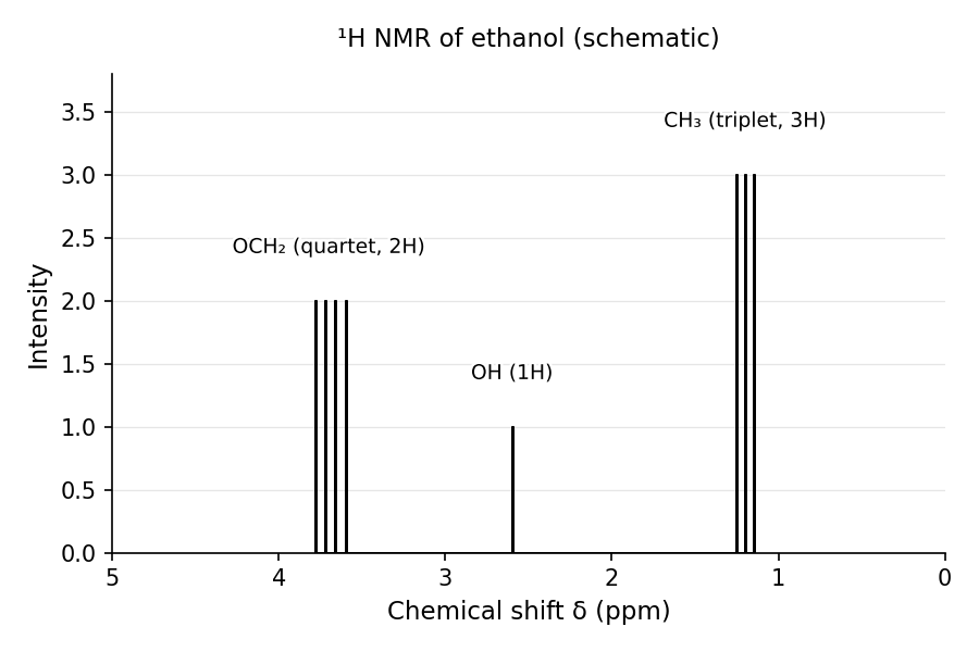
Must knowThe n + 1 rule: neighboring non-equivalent protons split each other's signal through bonds (vicinal, 2–3 bonds). A proton with adjacent non-equivalent protons gives lines, with intensities from Pascal's triangle:
| n | Pattern | Intensities |
|---|---|---|
| 0 | singlet | 1 |
| 1 | doublet | 1:1 |
| 2 | triplet | 1:2:1 |
| 3 | quartet | 1:3:3:1 |
The line spacing is the coupling constant (~6–8 Hz vicinal). Equivalent protons do NOT split each other (a CH's own H's don't split each other).
Worked Example — Ethanol (): CH (next to 2 H) → triplet; CH (next to 3 H) → quartet; OH → broad singlet (fast exchange suppresses coupling in protic solvents). This triplet + quartet is the canonical ethyl signature.
Integration ∝ number of protons giving a signal, so it gives the ratio of proton types—key for structure elucidation.
Quick check: In CHCHO, the CH is a doublet and the CHO is a quartet. Why? Answer: CH (3 H) is adjacent to 1 CHO proton → n+1 = doublet; CHO (1 H) is adjacent to 3 CH protons → n+1 = quartet. Each splits the other.
Common Confusions & Tricks
1. IR transmittance vs. absorbance: y-axis is transmittance, so peaks point downward; a dip means absorption.
2. Carbonyl wavenumber order: "Amides Are Always Low"—amide (~1650) < acid (~1710) < ketone/aldehyde (~1720) < ester (~1740). Ester is always higher than ketone.
3. Broad O–H vs. sharp N–H: both near 3300, but O–H (esp. carboxylic acid) is very broad from H-bonding. Primary amines show two N–H peaks, secondary one, tertiary none—a way to distinguish amine types.
4. UV-Vis: you see the complement. -Carotene absorbs blue-violet, so it looks orange, NOT blue. Always flip to the complementary color.
5. vs. : the band is always weaker and at longer wavelength. Of two carbonyl UV bands, the longer/weaker one is .
6. NMR: equivalent protons don't split each other. Benzene's six H's give a singlet; a CH's H's split only neighboring non-equivalent H's.
7. NMR: n+1 assumes equivalent neighbors. With non-equivalent neighbors in different environments you get complex multiplets; the MCAT tests only the clean cases.
8. Chemical shift direction: higher ppm = downfield = less shielded. Electronegative groups deshield → downfield. Don't reverse it.
9. IR vs. NMR roles: IR identifies what functional groups are present; NMR tells you how many and where (connectivity). Complementary tools.
10. Conjugation and color: more conjugation → smaller gap → longer absorbed (not shorter/bluer—a common reversal).
Key Equations
| Equation | Variables & Usage |
|---|---|
| Photon energy; absorption when matches a quantized transition. | |
| Wavenumber; = force constant, . Predicts IR shift with bond order or mass. | |
| Beer-Lambert; = molar absorptivity, = path length, = concentration. | |
| Absorbance vs. transmittance. ; . | |
| Chemical shift, referenced to TMS. Higher = downfield. | |
| Splitting | Lines in a multiplet = adjacent non-equivalent protons + 1. |
| Effective field on a proton; greater → upfield. |