Overview: Why This Topic Dominates the MCAT
Amino acids are the alphabet of biochemistry — every enzyme and structural protein is built from the same 20 building blocks. The MCAT tests this topic from several angles at once: organic chemistry (synthesis, stereochemistry), general chemistry (acid-base, isoelectric point), and biochemistry (structure-function).
Priority labels: Must know = cold; Know the logic = mechanism not names; Passage-level = recognize, don't memorize; Optional = skippable.
The organizing principle: the side chain (R group) determines everything — an amino acid's charge, its behavior in a protein, its isoelectric point, and how it folds.
Foundational Structure: A Recap
Must knowThe structure, stereochemistry, and acid-base behavior of amino acids are the foundational material covered in full in the 1A Amino Acids guide. This 5D guide is the organic-chemistry and analytical companion — it assumes that foundation and focuses on synthesis, peptide-bond reactions, and protein separation. The brief recap below orients you; for the complete treatment, see the 1A guide.
Structure. Every α-amino acid has a central α-carbon bonded to an amino group (), a carboxyl group (), a hydrogen, and a unique R group (side chain). Only the R group differs across the 20, so all share the same backbone acid-base chemistry and peptide-bond reactivity. At physiological pH the dominant form is the zwitterion .
Stereochemistry. All but glycine (R = H, achiral) have a chiral α-carbon. Biologically, amino acids are L ( on the left in a Fischer projection); most L-amino acids are also S by CIP. The classic trap: L-cysteine is R, because the sulfur in outranks the carboxyl in CIP priority even though the spatial arrangement matches every other L-amino acid.
Zwitterions and charge. The (acid) and (base) groups give a zwitterion with no net charge near neutral pH. At very low pH both protonate (net +1); at very high pH both deprotonate (net −1). Histidine () is the main amino-acid buffer near physiological pH.
Classification by side chain. Acidic (Asp, Glu — negative at pH 7); basic (Lys ~10.5, Arg ~12.5, His ~6); polar uncharged (Ser, Thr, Cys, Asn, Gln, Tyr); and nonpolar/hydrophobic (Gly, Ala, Val, Leu, Ile, Pro, Phe, Trp, Met), which drive the hydrophobic effect in the protein core. The high-yield standouts — glycine (achiral), proline (helix-breaker), cysteine (disulfides), histidine (buffer) — recur throughout this guide.
Synthesis of α-Amino Acids
Know the logic of two named syntheses — what they start from and produce — not every intermediate.
Strecker Synthesis
Know the logicBuilds an α-amino acid from an aldehyde. An aldehyde () reacts with (from ) to form an imine, which attacks to give an α-aminonitrile; hydrolysis of the nitrile yields the carboxylic acid.
Remember: the aldehyde carbon becomes the α-carbon; cyanide is the nucleophile and becomes the carboxyl; product is racemic.
Quick check: In Strecker synthesis of alanine (R = methyl), what aldehyde would you start with?
Answer: Acetaldehyde (); its carbonyl carbon becomes the α-carbon of alanine.
Gabriel Synthesis
Know the logicUses the phthalimide anion as a protected form of . Because phthalimide's nitrogen sits between two carbonyls, it is poorly nucleophilic — so alkylation stops cleanly at a primary amine (no over-alkylation). You alkylate phthalimide (often via a malonic-ester route to add the carboxyl), then hydrolyze off the phthalimide (acid, base, or hydrazine) to release the free . Product is racemic.
Quick check: Why is Gabriel synthesis preferred over direct alkylation of ammonia?
Answer: Direct alkylation of gives mixtures of 1°, 2°, 3° amines and quaternary salts because each product is at least as nucleophilic as . Phthalimide's nitrogen is far less nucleophilic (flanked by two carbonyls), ensuring a single, primary-amine product.
Isoelectric Point and Titration: A Recap
Must knowThe isoelectric point (pI) — the pH of zero net charge, where the molecule doesn't migrate in a field — and the reading of amino-acid titration curves are covered in full in the 1A Amino Acids guide. They are recapped briefly here because they underpin the separation techniques (electrophoresis, isoelectric focusing) that this guide develops.
The pI is the average of the two pKa values that flank the neutral (zwitterionic) form:
For a simple amino acid, average the carboxyl (~2) and amino (~9–10) pKa's. For an acidic AA (Asp, Glu) average the two carboxyl pKa's → pI ≈ 3; for a basic AA (Lys, Arg, His) average the two amino pKa's → pI > 7. On a titration curve, each flat buffering plateau is centered on a pKa (half-protonated, pH = pKa) and the pI is the flat midpoint between the two pKa's flanking the neutral species. See the 1A guide for worked examples.
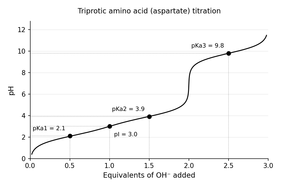
Quick check: For lysine (; α-amino; ε-amino), what is the pI?
Answer: The neutral zwitterion lies between the two amino pKa's: . Above 7, as expected for a basic amino acid.
Peptides and Proteins: Reactions
Peptide Linkage
Must knowWhen two amino acids join, the carboxyl of one and the amino of the next undergo a condensation (dehydration), releasing water and forming an amide (peptide) bond .
Key features:
- Partial double-bond character: nitrogen's lone pair delocalizes into the carbonyl, making the bond planar with restricted rotation.
- Trans preference: the two α-carbons adopt trans to minimize steric clash (proline is the exception).
- Directionality: chains run N-terminus (free ) → C-terminus (free ), written N→C, left to right.
- Counting: residues → peptide bonds.
Quick check: A polypeptide contains 100 residues. How many peptide bonds, and how many water molecules were released during synthesis?
Answer: 99 peptide bonds; 99 water molecules (one per bond formed).
Sulfur Linkage: Cysteine and Cystine
Must knowTwo cysteine thiols () — in the same or different chains — oxidize to a covalent disulfide bond ():
The oxidized, disulfide-linked pair is called cystine (distinct from cysteine). Reducing agents (β-mercaptoethanol, DTT) cleave it back to free thiols.
- Disulfides are covalent — the strongest bonds in tertiary/quaternary structure.
- They form mainly in extracellular proteins or the ER lumen (oxidizing environments); the reducing cytoplasm keeps most cysteines as free .
Quick check: A biochemist adds DTT (a reducing agent) to purified insulin (two chains held partly by disulfides). What happens?
Answer: DTT reduces the disulfides (), so insulin's two chains separate — showing the disulfides stabilize the chains' association.
Hydrolysis of Peptides and Proteins
Must knowHydrolysis adds water across the amide bond (the reverse of formation):
It can be acid-catalyzed, base-catalyzed, or enzymatic (proteases with residue specificity):
- Trypsin: cleaves after basic residues Lys (K), Arg (R).
- Chymotrypsin: cleaves after large aromatic residues (Phe, Trp, Tyr).
- Pepsin: stomach (low pH); cleaves after aromatic/bulky residues.
(Edman degradation is beyond MCAT scope — just know proteases cleave at specific residues.)
Separating and Analyzing Proteins
Passage-levelRecognize what each technique separates by:
- Gel electrophoresis: smaller/more-charged migrate faster.
- SDS-PAGE: uniform negative coating → separates by size alone (estimates MW).
- Isoelectric focusing (IEF): each protein stops at the pH = its pI.
- Ion-exchange: by charge. Size-exclusion: by size (large molecules elute first).
Quick check: A polypeptide ARKFGDW is treated with trypsin. What fragments result?
Answer: Trypsin cleaves after K and R: after R (position 2) → AR; after K (position 3) → K; the rest FGDW has no internal K/R. Fragments: AR, K, FGDW.
Protein Structure: General Principles and the Four Levels
Must knowA protein's function depends on its 3D shape, organized into four hierarchical levels, each stabilized by different interactions.
Primary Structure
Must knowPrimary structure is the linear sequence of amino acids, joined by covalent peptide bonds and dictated by the DNA sequence. It determines all higher levels — the sequence contains all folding information. The only covalent bonds here are peptide bonds (and disulfides, which depend on primary structure to position the cysteines).
Quick check: Sickle-cell anemia results from a single change in β-globin: glutamate (position 6) → valine. At what level of structure is this mutation?
Answer: Primary structure — a change in sequence. This one covalent change cascades upward: a surface that should be hydrophilic becomes hydrophobic, causing tetramer aggregation under low oxygen.
Secondary Structure
Must knowSecondary structure is local, regular, repeating backbone hydrogen bonding — between the of one peptide bond and the of another. R groups do not participate.
α-Helix: a right-handed coil stabilized by intrastrand H-bonds between residue () and residue (); side chains point outward (~3.6 residues per turn). Proline disrupts it (no to donate; rigid kink).
β-Sheet: extended strands H-bond interstrand. Antiparallel (strands opposite N→C) has more linear, stronger H-bonds → more stable than parallel. Side chains alternate above/below the plane.
β-Turn: a tight 4-residue reversal stabilized by one H-bond; commonly contains proline and glycine.
Quick check: Why can't proline be in the middle of an α-helix?
Answer: Proline's nitrogen is locked in a ring with no to donate the backbone H-bond that stabilizes the helix; the ring also constrains the backbone into a rigid kink.
Tertiary Structure
Must knowTertiary structure is the complete 3D fold of a single chain, arising from R-group interactions: hydrophobic effect, hydrogen bonds, ionic interactions / salt bridges (Asp/Glu with Lys/Arg), disulfide bonds, and van der Waals forces.
The hydrophobic effect is the dominant driving force: burying nonpolar residues releases ordered water into the bulk, increasing entropy — so folding is primarily entropy-driven.
Denaturation disrupts secondary/tertiary structure without breaking peptide bonds. Agents: urea/guanidinium (break H-bonds and hydrophobic packing), heat, detergents (SDS), reducing agents (β-mercaptoethanol/DTT, for disulfides), and extreme pH (disrupts salt bridges).
Quick check: A membrane protein has a transmembrane domain passing through the hydrophobic lipid bilayer. Predict its composition.
Answer: The transmembrane segment is rich in nonpolar/hydrophobic residues (Val, Leu, Ile, Ala, Phe) so the side chains pack favorably with the lipid acyl chains; charged/polar residues there are unfavorable unless functionally essential.
Quaternary Structure
Must knowQuaternary structure is the assembly of two or more subunits into one complex, held by the same noncovalent forces as tertiary structure (plus occasional disulfides). Only multi-subunit proteins have it.
The canonical example is hemoglobin, an tetramer whose subunits cooperate: binding to one raises the others' affinity (cooperativity, sigmoidal curve). This is why the surface Glu→Val sickle mutation matters at the quaternary level — deoxygenated tetramers aggregate into fibers.
Fibrous vs. Globular Proteins
Must knowGlobular proteins (hemoglobin, most enzymes) fold into compact, roughly spherical, usually water-soluble shapes with hydrophobic cores. Fibrous proteins are elongated, strong, and typically insoluble (structural roles):
- Collagen: a triple helix on a repeating Gly-X-Y sequence; glycine at every third position is essential (only its tiny H side chain fits the crowded interior). Hydroxyproline (needs vitamin C) stabilizes it — deficiency causes scurvy.
- Keratin: α-helical fibrous protein (hair, nails) rich in cysteine, cross-linked by disulfides.
Common Confusions & Tricks
1. Cysteine is L but R — know this cold. Same spatial arrangement as other L-amino acids, but CIP designation is R because the side chain's sulfur has high priority. "All L-amino acids are S" is false.
2. Cysteine (thiol) vs. Cystine (disulfide). Cysteine = monomer with free ; cystine = the oxidized, disulfide-linked dimer.
3. pI: average the two pKa's flanking the neutral species. The common error is averaging ALL pKa's. Acidic AAs: both carboxyls. Basic AAs: both nitrogens.
4. At pH = pKa, the species is 50% protonated / 50% deprotonated — not at its pI. Henderson-Hasselbalch: pH = pKa means [A] = [HA].
5. N-terminus vs. C-terminus. Sequences written N→C (left to right). N-terminus = free ; C-terminus = free .
6. Hydrolysis vs. Denaturation. Denaturation unfolds secondary/tertiary structure but leaves peptide bonds intact; hydrolysis breaks peptide bonds. SDS-PAGE denatures; proteases hydrolyze.
7. Antiparallel β-sheet is more stable than parallel. Antiparallel H-bonds are more linear and thus stronger.
8. Strecker = aldehyde + cyanide + hydrolysis. The aldehyde carbon becomes the α-carbon; cyanide adds the nitrogen-adjacent carbon that becomes the carboxyl after hydrolysis.
9. Histidine is the buffer at physiological pH. Its side-chain pKa (~6) is the only one in the physiological range — key in enzyme active sites and hemoglobin (Bohr effect).
10. Proline disrupts α-helices; glycine is flexible — both appear in β-turns. Proline can't donate an H-bond; glycine has no R group. Neither forms good extended secondary structure.
Key Equations
| Equation | Variables and Use |
|---|---|
| Use the two pKa's flanking the neutral (zero-charge) species. Simple AAs: α-COOH and α-NH. Acidic AAs: both COOH. Basic AAs: both amino groups. | |
| Henderson-Hasselbalch. Predicts charge state at a given pH. At : 50% protonated, 50% deprotonated. | |
| Peptide bonds: | A polypeptide of residues has peptide bonds and released water molecules during biosynthesis. |
| Oxidation of two cysteine thiols to a disulfide (cystine); reversed by DTT / -mercaptoethanol. | |
| Hydrolysis of a peptide bond. Trypsin: after K, R; chymotrypsin: after F, W, Y. |