Proteins do far more than catalyze reactions. Much of cellular life depends on proteins that bind, signal, defend, and move — all without acting as enzymes. The MCAT tests these roles heavily: hemoglobin's cooperative binding, antibodies, and ATP-powered motor proteins.
Priority labels: Must know = cold; Know the logic = mechanism not names; Passage-level = recognize, don't memorize; Optional = skippable.
Ligand Binding by Nonenzymatic Proteins
The Core Concept
Must knowA ligand is any molecule that binds specifically and reversibly to a protein at a binding site complementary to it in shape and charge. Binding can be independent (one ligand per site, no communication) or cooperative (binding at one site changes affinity at another). Hemoglobin is the canonical cooperative example.
Myoglobin vs. Hemoglobin
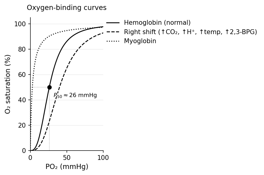
Must knowMyoglobin (Mb) is a monomeric O₂-storage protein in muscle. With only one heme, there is no cooperativity, so its O₂-binding curve is a hyperbola:
where is fractional saturation. Myoglobin has high O₂ affinity () — it holds O₂ tightly and releases it only at very low , ideal for storage.
Hemoglobin (Hb) is a tetramer (two α + two β subunits, four hemes). The subunits communicate: binding the first O₂ makes the next bind more easily — positive cooperativity — giving a sigmoidal (S-shaped) curve.
In the lungs ( mmHg) Hb is ~98% saturated; in tissues ( mmHg) it drops to ~75%, unloading O₂. The steep middle means small changes produce large changes in O₂ delivery — exactly what a transport molecule needs.
The T State and R State
Know the logicHemoglobin toggles between two conformations — T (Tense), low-affinity, deoxy-Hb; and R (Relaxed), high-affinity, oxy-Hb. As O₂ binds, the equilibrium shifts toward R, raising affinity for the next O₂. This is the molecular basis of cooperativity and the sigmoid. Reason about cooperativity qualitatively — no Hill equation needed.
( at 50% saturation) measures affinity; normal Hb mmHg.
Allosteric Modulators and the Bohr Effect
Must knowRight shift (↑, lower affinity, more O₂ unloading): ↑, ↑temperature, ↑2,3-BPG, ↓pH (↑[H⁺]).
Left shift (↑ affinity, less unloading): the opposites — plus carbon monoxide (CO), which binds Hb ~200× more tightly than O₂ and locks it in the R state.
The Bohr effect is the right shift from CO₂ and H⁺: active tissues accumulate CO₂/H⁺, lowering pH and right-shifting the curve to release O₂ where needed; the lungs reverse this.
2,3-BPG is a glycolytic intermediate that rises in RBCs under hypoxia (high altitude, anemia). It binds and stabilizes the T state, right-shifting the curve to deliver more O₂.
CO₂ Transport and the Chloride Shift
Know the logicHb also carries CO₂ to the lungs. A small fraction binds globin N-termini as carbaminohemoglobin (stabilizes T, aiding the Bohr effect). Most CO₂ is converted by carbonic anhydrase: . The is buffered by Hb (driving O₂ release); is exported in exchange for entering — the chloride shift. The lungs reverse the whole process.
Fetal Hemoglobin (HbF)
Must knowHbF has two γ-subunits instead of β. γ binds 2,3-BPG poorly, so HbF has higher O₂ affinity (left-shifted vs. adult HbA) — letting the fetus pull O₂ from maternal blood across the placenta.
Other Binding Proteins
Passage-levelRecognize a few non-Hb binding proteins — albumin (carries fatty acids, hormones, drugs in blood), transferrin/ferritin (iron transport/storage), calmodulin (Ca²⁺ sensor), G proteins (GTP/GDP switch), nuclear receptors (steroid hormones → transcription).
Cell-surface receptors bind an extracellular ligand and transduce a signal across the membrane without chemically transforming it. GPCRs activate intracellular G proteins; receptor tyrosine kinases (RTKs) (e.g., insulin receptor) dimerize on ligand binding. The recognition event itself is pure binding — the defining nonenzymatic function.
Quick check: A climber spends several weeks at high altitude. Which direction does her hemoglobin O₂ dissociation curve shift, and what drives it?
Answer: Right shift. Chronic hypoxia increases 2,3-BPG, which stabilizes the T state of deoxy-Hb, lowering O₂ affinity and promoting O₂ delivery despite lower arterial .
Antibody–antigen binding is itself a high-specificity, non-catalytic recognition/binding event; that chemistry, along with the immune system (innate/adaptive immunity, antibody structure, MHC, clonal selection, isotypes, active vs. passive immunity), is covered in the 1A Nonenzymatic Protein Function guide.
Motor Proteins
Must knowMotor proteins are mechanochemical transducers: they couple ATP hydrolysis (, ) to conformational changes that produce mechanical work (force × displacement) rather than chemically transforming a substrate. The key examples are myosin (walks along actin to drive muscle contraction) and kinesin and dynein (walk along microtubules to transport cargo). The detailed sliding-filament/cross-bridge mechanism and kinesin–dynein transport are covered in the 1A Nonenzymatic Protein Function guide.
Common Confusions & Tricks
1. Left vs. right shift — ask "does this help or hinder O₂ delivery?" Acidosis, fever, exercise, high altitude → right shift (unload more O₂). Cold, alkalosis, CO → left shift (hold tighter). Reason from physiology: exercising muscle is hot, acidic, high-CO₂ → needs O₂ → right-shift.
2. Fetal Hb is left-shifted (HIGHER affinity) — so it can steal O₂ from maternal HbA across the placenta. Not "lower affinity."
3. The antibody binds; it does not catalyze. Specificity is in the variable/CDR regions; the Fc region recruits effectors. Don't call it an "enzyme that destroys antigen."
4. A-band does NOT change during contraction. Only the sarcomere, I-band, and H-zone shorten. A-band = thick-filament length = constant.
5. ATP releases myosin from actin (step 1), NOT the power stroke. ATP hydrolysis cocks the head; Pi release powers the stroke. Rigor mortis = no ATP = stuck.
6. Kinesin ≠ dynein direction: Kinesin = out (anterograde, + end); dynein = in (retrograde, − end). Cilia use axonemal dynein; Kartagener = dynein defect.
7. 2,3-BPG stabilizes the T state, not the R state. High altitude → ↑2,3-BPG → T stabilized → right shift → more O₂ delivery.
8. Reason about cooperativity qualitatively. Sigmoidal = cooperative (hemoglobin); hyperbolic = non-cooperative (myoglobin). No Hill coefficient required.
Key Relationships
Conceptual relationships, not equations to compute (no binding-curve math at MCAT scope).
| Concept | What to know |
|---|---|
| Lower = tighter binding (higher affinity). | |
| Binding-curve shape | Hyperbolic = single-site, non-cooperative (myoglobin); sigmoidal = multi-subunit, cooperative (hemoglobin). |
| at 50% saturation. ↑ = right shift (↓ affinity, more unloading). Normal Hb ≈ 26 mmHg. | |
| Motor proteins & ATP () | One ATP per myosin cross-bridge cycle; one ATP per kinesin step. |
| Sarcomere geometry | A-band constant during contraction; I-band and H-zone shorten. |