Sound lets you detect threats, communicate, and navigate space without light. The MCAT tests hearing both structurally (the anatomical compartments and what each does) and functionally (how a pressure wave in air becomes a conscious auditory experience). This guide builds from the outside of the ear inward to the temporal lobe.
Priority labels: Must know = cold; Know the logic = mechanism not names; Passage-level = recognize, don't memorize; Optional = skippable.
Structure and Function of the Ear
The Outer Ear: Capturing Sound
Must knowThe outer ear consists of the pinna (the visible cartilaginous flap) and the external auditory canal, which funnel sound toward the eardrum. The pinna's shape aids sound localization, especially front-vs-back. The canal ends at the tympanic membrane (eardrum), which vibrates in response to the compressions and rarefactions of a sound wave. The outer ear simply captures and directs — the whole ear's job is to convert mechanical vibration in air into an electrical signal the brain can use.
Sound as a stimulus: Sound is a longitudinal pressure wave. Its frequency (Hz) determines perceived pitch (higher frequency = higher pitch); its amplitude determines perceived loudness; waveform complexity determines timbre. The healthy human ear detects roughly 20 Hz to 20,000 Hz, and high-frequency sensitivity declines with age (presbycusis).
Quick check: Why is the tympanic membrane cone-shaped rather than flat?
Answer: The cone shape increases sensitivity and couples sound energy efficiently into the ossicles across a range of frequencies.
The Middle Ear: Impedance Matching
Must knowSound travels in air, but the inner ear is fluid-filled. Fluid has much higher impedance than air, so most sound energy would reflect off the air-fluid interface without amplification. The middle ear matches impedance between airborne sound and inner-ear fluid.
Three tiny bones, the ossicles, span the middle-ear cavity: malleus (attached to the eardrum) → incus → stapes (footplate presses on the oval window). They amplify mechanically via (1) lever action of the ossicle chain and (2) the area ratio — the large eardrum concentrating force onto the much smaller oval window — together giving a large pressure boost needed to cross the air-to-fluid barrier.
The oval window is the input where the stapes transmits vibrations into the inner ear. The round window bulges outward to let the incompressible cochlear fluid move when the oval window is pushed in.
The Eustachian tube connects the middle ear to the nasopharynx and opens during swallowing/yawning to equalize pressure across the eardrum ("ear pop"). Passage-level — chronic Eustachian dysfunction can cause otitis media (middle-ear fluid/infection), a classic context for conductive hearing loss.
Quick check: A patient's stapes has fused to the oval window (otosclerosis). What type of hearing loss results, and why?
Answer: Conductive hearing loss. The stapes can no longer vibrate to transmit sound inward, even though the cochlear hair cells are intact.
The Inner Ear: Transduction Begins
Must knowThe inner ear contains the cochlea (hearing) and the vestibular system (balance); this guide focuses on the cochlea.
The cochlea is a snail-shaped, fluid-filled structure with three chambers running its length:
| Chamber | Fluid | Connects to |
|---|---|---|
| Scala vestibuli | Perilymph | Oval window |
| Scala media (cochlear duct) | Endolymph | enclosed |
| Scala tympani | Perilymph | Round window |
Perilymph resembles extracellular fluid (high Na⁺, low K⁺). Endolymph is the unusual one — high K⁺, low Na⁺ (resembling intracellular fluid). This composition is what powers hair-cell transduction. The scala vestibuli and scala tympani join at the helicotrema at the apex.
The scala media is bounded by Reissner's membrane above and the basilar membrane below. Resting on the basilar membrane is the organ of Corti, which holds the sensory hair cells. Stapes movement pushes the oval window → fluid displacement travels along the scala vestibuli → the basilar membrane waves up and down → the round window bulges to accommodate.
Quick check: If a tumor destroyed the round window membrane, what would happen to hearing?
Answer: Hearing would be severely impaired. Without the round window bulging outward, the incompressible cochlear fluid cannot move, so the basilar membrane cannot deflect and hair cells cannot be stimulated.
Place Theory and Tonotopy: Frequency Encoding on the Basilar Membrane
Must knowPlace theory (Georg von Békésy's traveling wave) explains how the cochlea distinguishes pitch. The basilar membrane varies along its length:
- Base (near oval window): narrow, stiff → resonates best at high frequencies
- Apex (helicotrema): wide, floppy → resonates best at low frequencies
A pure tone causes maximum basilar-membrane displacement at a specific location — tonotopic organization. The brain identifies which frequencies are present by which hair cells fire (place/spatial coding).
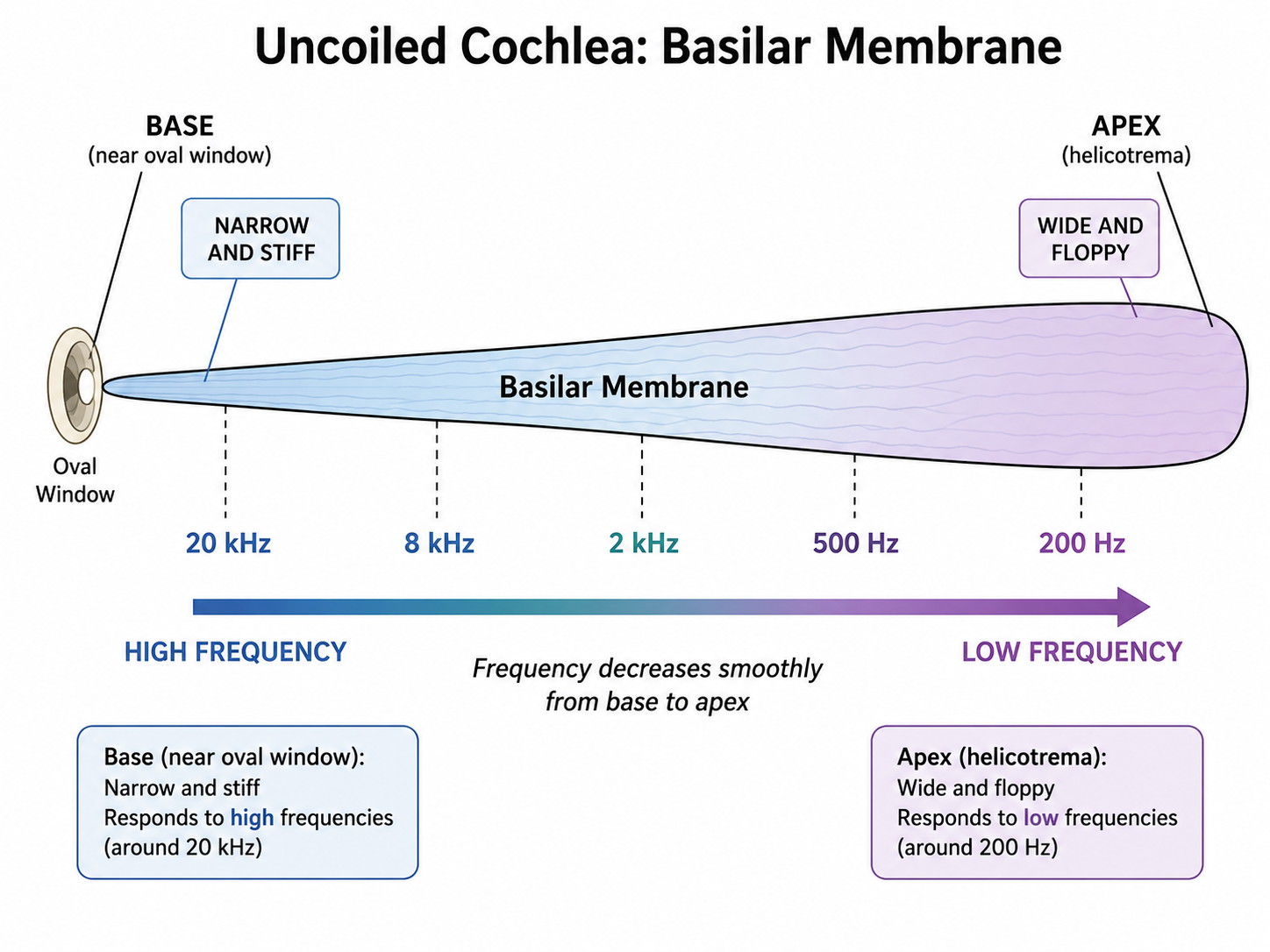
Place theory falters at low frequencies, where membrane-position differences become too subtle. Temporal (frequency) theory holds that the auditory nerve fires in synchrony with the sound wave, so firing rate encodes low frequencies. The volley principle extends this: groups of neurons take turns firing so they collectively track frequencies too fast for any single neuron. Loudness is coded by how many hair cells fire and how fast. Know the logic: know that both theories exist and what each explains — place for high frequencies, temporal/volley for low.
Quick check (scenario): A patient has cochlear damage at the base of the basilar membrane. What hearing loss would you predict?
Answer: Loss of high-frequency sounds, since the base resonates at high frequencies. This is the pattern in noise-induced hearing loss, which disproportionately damages the basal region.
Sensory Reception by Hair Cells
Hair Cell Anatomy and the Organ of Corti
Must knowThe organ of Corti sits on the basilar membrane and contains two cell types:
- Inner hair cells: the true sensory receptors; almost all auditory nerve fibers synapse here.
- Outer hair cells: amplifiers that actively contract (via the protein prestin) to boost basilar-membrane movement and sharpen frequency selectivity.
Each hair cell is topped with a staircase of stiff stereocilia. The tallest stereocilia contact the overlying tectorial membrane, so when the basilar membrane moves, the shearing force between the two membranes deflects the stereocilia.
Mechanotransduction: How Deflection Becomes an Electrical Signal
Must knowThe core mechanism:
- Deflection toward the tallest stereocilia opens mechanically gated K⁺ channels at the stereocilia tips.
- K⁺ flows INTO the hair cell from the endolymph. This is counterintuitive, but endolymph is uniquely high in K⁺ and the cell interior is negative, so K⁺ enters down both its concentration and electrical gradients.
- Depolarization opens voltage-gated Ca²⁺ channels at the base → Ca²⁺ influx → vesicle fusion → glutamate release onto the cochlear branch of cranial nerve VIII (vestibulocochlear nerve).
- Deflection the other way closes the channels → hyperpolarization → less neurotransmitter.
The hair cell is a mechano-electric converter: mechanical deflection → ionic current → chemical synapse → action potential in the auditory nerve.
Quick check (scenario): Aminoglycoside antibiotics (e.g., gentamicin) destroy cochlear hair cells. Conductive or sensorineural loss, and why?
Answer: Sensorineural. The damage is to the sensory receptor cells, not the mechanical conduction pathway. It is generally irreversible because mammalian hair cells do not regenerate.
Why the Endolymph Matters
Know the logicThe stria vascularis (lateral wall of the scala media) pumps K⁺ into the endolymph, keeping it both high in K⁺ and electrically positive. This creates a large driving force pushing K⁺ into hair cells the instant their channels open, making them exquisitely sensitive to tiny deflections. (Exact membrane-potential values are beyond MCAT scope.)
Auditory Processing (Auditory Pathways in the Brain)
From Cochlea to Cortex: The Ascending Pathway
Must knowUnlike vision, the auditory pathway has extensive brainstem processing and is bilateral — sound from one ear is processed by both hemispheres, so unilateral cortical lesions rarely cause one-ear deafness.
The ascending pathway: cochlear nerve (CN VIII) → cochlear nucleus → superior olivary complex → inferior colliculus → medial geniculate nucleus (thalamus) → primary auditory cortex (temporal lobe). Two stations carry the highest-yield functions: the superior olivary complex (SOC) does binaural sound localization, and the medial geniculate nucleus (MGN) is the thalamic relay to cortex. Optional handle — "Sound Climbs Slowly, Like Intelligent Monkeys" (the seven stations in order).
Tonotopic organization is preserved at every level, not just the cochlea.
Quick check (scenario): A patient has a stroke in the left inferior colliculus. Would you expect them to be deaf in the right ear?
Answer: No. By the inferior colliculus, signals are already bilaterally represented, so a unilateral lesion at this level or above does not cause complete contralateral deafness (though subtle localization deficits may appear).
The Auditory Cortex and Higher Processing
Must knowPrimary auditory cortex (A1) (Heschl's gyri of the superior temporal gyrus) does initial cortical analysis of pitch, rhythm, and onset, and is tonotopically mapped.
Wernicke's area (posterior superior temporal gyrus, usually left hemisphere) is critical for language comprehension; damage causes Wernicke's aphasia (fluent but meaningless speech, poor comprehension). Contrast with Broca's area (left inferior frontal gyrus → speech production).
Sound localization (two cues):
- Interaural time difference (ITD): sound reaches the near ear first; used for low frequencies.
- Interaural level difference (ILD): the head's "sound shadow" makes the far ear receive softer sound; used for high frequencies.
Quick check (scenario): A sound directly in front and one directly behind produce equal ITDs and ILDs. How does the auditory system tell front from back?
Answer: The pinna filters sound frequency-specifically (head-related transfer functions) differently for front vs. behind; the brain has learned these spectral cues. This is why the pinna's shape matters.
Hearing Loss: Types and MCAT Context
Must knowLocate the lesion:
Conductive hearing loss: mechanical problem in the outer or middle ear (cerumen impaction, ruptured eardrum, otosclerosis, otitis media). Hair cells intact; often treatable.
Sensorineural hearing loss: problem in the cochlear hair cells or auditory nerve (noise exposure — basal outer hair cells first; aminoglycosides; presbycusis; acoustic neuroma on CN VIII). Generally permanent, since hair cells don't regenerate.
Mixed hearing loss combines both.
Quick check (scenario): Normal outer/middle ear and intact ossicle movement, but reduced high-frequency hearing after years near loud machinery. Conductive or sensorineural?
Answer: Sensorineural. The conduction pathway is intact; the damage is to cochlear hair cells (noise preferentially harms basal, high-frequency outer hair cells).
Common Confusions & Tricks
1. Endolymph ≠ ordinary extracellular fluid. Endolymph is high K⁺, low Na⁺ (opposite of typical extracellular fluid). This is why K⁺ flows into hair cells during transduction. See endolymph → think "high K⁺."
2. Round window vs. oval window. Oval = INPUT (stapes presses on it). Round = OUTPUT (pressure relief). Mnemonic: Oval = Ossicle input; Round = Relief.
3. Base vs. apex frequency — often reversed. The BASE (near the oval window) responds to HIGH frequencies. Anchor it: the base is stiff and narrow → stiff things vibrate fast → high frequency.
4. Inner vs. outer hair cells. Inner = primary sensory receptors (~95% of afferent fibers). Outer = amplifiers (electromotility via prestin). If asked which are the primary receptors, answer inner.
5. Auditory pathway laterality trap. The pathway is bilateral at every level above the cochlear nucleus. A left-hemisphere lesion does not cause right-ear deafness. Only a lesion at the cochlea or CN VIII produces ipsilateral deafness.
6. Sensorineural vs. conductive — locate the lesion. Anything before the oval window (earwax, ruptured eardrum, otosclerosis, middle-ear fluid) with intact hair cells → conductive. Damage to the cochlea or CN VIII (noise, ototoxic drugs, aging, acoustic neuroma) → sensorineural, generally permanent.
7. Auditory relay stations. Order: cochlear nucleus → superior olive → inferior colliculus → medial geniculate → cortex. Don't confuse inferior colliculus (auditory) with superior colliculus (visual) — both in the midbrain tectum.
8. Wernicke's vs. Broca's. Wernicke's = comprehension, posterior temporal. Broca's = production, frontal. Trick: Wernicke's = What did you say? Broca's = Being able to speak.
Key Theories & Terms
| Term / Name | One-sentence Summary |
|---|---|
| Pinna | Outer cartilaginous flap that funnels sound into the ear canal and aids front-back localization. |
| Tympanic membrane | The eardrum; vibrates in response to sound and couples it to the ossicles. |
| Ossicles (malleus, incus, stapes) | Three middle-ear bones that amplify and transmit vibrations from eardrum to oval window. |
| Eustachian tube | Canal connecting middle ear to nasopharynx; equalizes pressure across the eardrum. |
| Oval window | Membrane where the stapes delivers vibrations to the inner ear (input). |
| Round window | Membrane that bulges outward to allow cochlear fluid movement (pressure relief). |
| Cochlea | Fluid-filled, coiled inner-ear structure housing the organ of Corti; performs frequency analysis. |
| Scala vestibuli / scala tympani | Perilymph-filled chambers flanking the scala media; carry pressure waves from oval to round window via the helicotrema. |
| Scala media (cochlear duct) | Endolymph-filled chamber housing the organ of Corti. |
| Perilymph | High-Na⁺, low-K⁺ fluid in scala vestibuli/tympani; like extracellular fluid. |
| Endolymph | High-K⁺, low-Na⁺ fluid in the scala media; its composition drives hair-cell transduction. |
| Basilar membrane | Narrow/stiff at base (high-frequency), wide/floppy at apex (low-frequency). |
| Tectorial membrane | Overlies the stereocilia; shearing against the basilar membrane deflects them. |
| Organ of Corti | Sensory epithelium on the basilar membrane containing inner and outer hair cells. |
| Inner hair cells | Primary auditory receptors; receive ~95% of afferent cochlear fibers. |
| Outer hair cells | Electromotile amplifiers; use prestin to amplify basilar-membrane motion. |
| Stereocilia | Stiff projections whose mechanically gated K⁺ channels open on deflection toward the tall side. |
| Mechanotransduction | Conversion of mechanical stereocilia deflection into an electrical signal. |
| Stria vascularis | Lateral wall of the scala media; pumps K⁺ into endolymph to power transduction. |
| Place theory (Békésy) | Pitch encoded by the location of maximal basilar-membrane displacement; best for high frequencies. |
| Temporal (frequency) theory | Auditory nerve firing rate encodes frequency; applies to low frequencies. |
| Volley principle | Groups of neurons fire in rotating volleys to encode frequencies too high for one neuron. |
| Tonotopy | Ordered frequency mapping preserved from cochlea to cortex. |
| Cochlear nucleus | First brainstem relay of CN VIII; signals diverge to both sides here. |
| Superior olivary complex (SOC) | Pontine nucleus for binaural processing and localization via ITD/ILD. |
| Inferior colliculus | Midbrain auditory integration center; mediates reflexive orienting/startle. |
| Medial geniculate nucleus (MGN) | Thalamic relay for auditory signals en route to cortex. |
| Primary auditory cortex (A1) | Heschl's gyri of the temporal lobe; tonotopically organized first cortical processing. |
| Wernicke's area | Posterior superior temporal gyrus (left); language comprehension; damage causes Wernicke's aphasia. |
| Conductive hearing loss | Impaired mechanical transmission through outer/middle ear. |
| Sensorineural hearing loss | Damage to cochlear hair cells or auditory nerve; typically permanent. |
| Interaural time difference (ITD) | Arrival-time difference between ears; low-frequency localization. |
| Interaural level difference (ILD) | Intensity difference from head shadowing; high-frequency localization. |
| Prestin | Motor protein in outer hair cells responsible for electromotility/amplification. |