Vision is one of the most thoroughly tested sensory systems on the MCAT. It connects optics, cellular signaling, and perceptual psychology in one topic. Focus on understanding why each structure or mechanism exists rather than memorizing it.
Priority labels: Must know = cold; Know the logic = mechanism not names; Passage-level = recognize, don't memorize; Optional = skippable.
Structure and Function of the Eye
The Eye as an Optical Instrument
Must knowThink of your eye as a biological camera: light enters, gets focused onto a photosensitive surface, and a signal is sent to the brain. The light path is cornea → pupil → lens → vitreous → retina.
The cornea is the clear, curved front surface and provides most of the eye's refractive power (more than the lens), because the largest change in refractive index is at the air–cornea interface. The iris is the colored muscular ring that controls the diameter of the pupil (the aperture) — dilating in dim light (sympathetic) and constricting in bright light (parasympathetic).
The lens fine-tunes focus through accommodation: the ciliary muscles contract, releasing tension on the zonular fibers (suspensory ligaments), allowing the elastic lens to round up and increase its refractive power for near vision. When the ciliary muscles relax, the lens flattens for distance. Note the counterintuitive relationship — contracted ciliary muscle = rounder lens = near focus.
The vitreous humor behind the lens maintains the eye's shape. (The aqueous humor in front maintains intraocular pressure.)
The Retina: Where Light Becomes Signals
Must knowThe retina lines the back of the eye and is the neural tissue where phototransduction occurs.
| Feature | Rods | Cones |
|---|---|---|
| Light sensitivity | High (scotopic = dim/night) | Lower (photopic = bright) |
| Location | Peripheral retina | Concentrated at fovea |
| Color | No (one pigment: rhodopsin) | Yes (three types: S, M, L) |
| Acuity | Low (high convergence) | High (near 1:1 at fovea) |
The fovea is the central pit, packed with cones, where acuity is highest. The optic disc (blind spot) is where the optic nerve exits — no photoreceptors, so nothing is detected there.
Signal flow: photoreceptors → bipolar cells → retinal ganglion cells (RGCs) → optic nerve. (Horizontal and amacrine cells modulate signals laterally — off-scope detail.)
Refractive Errors and Corrective Lenses
Must know- Myopia (nearsightedness): eyeball too long, focal point falls in front of the retina; distant objects blurry. Corrected with a diverging (concave) lens.
- Hyperopia (farsightedness): eyeball too short, focal point falls behind the retina; near objects blurry. Corrected with a converging (convex) lens.
- Presbyopia: age-related loss of lens elasticity → reduced accommodation for near vision. Corrected with converging reading lenses.
- Passage-level Astigmatism — uneven corneal curvature focuses light at multiple points; cylindrical lens.
Quick check: A person with myopia needs which type of corrective lens, and why?
Answer: A diverging (concave) lens. In myopia, light converges too early (in front of the retina), so you need a lens that first diverges the light to push the focal point back onto the retina.
Visual Processing: Phototransduction and Color Vision
Phototransduction — The Signal Is a Hyperpolarization
Must knowUnlike most sensory neurons, photoreceptors are depolarized in the dark and hyperpolarize in response to light.
In the dark, a steady inward current — the "dark current" — keeps the cell depolarized and continuously releasing glutamate. When light strikes the photopigment (11-cis retinal bound to rhodopsin in rods), a cascade closes the dark-current channels, so the photoreceptor hyperpolarizes and reduces glutamate release; bipolar cells read this drop as the "light signal."
Photoreceptors do not fire all-or-none action potentials — they respond with a graded receptor potential. (The molecular cascade — transducin, PDE, cGMP — is off-scope; do not memorize it.)
Trichromatic Theory and Color Vision
Must knowThe trichromatic (Young–Helmholtz) theory: color vision is based on three cone types, each maximally sensitive to short (S, blue), medium (M, green), or long (L, red) wavelengths. The brain perceives color by comparing the ratio of activation across the three (e.g., red + green light → yellow).
Color blindness is most commonly an X-linked recessive condition in which one cone type is absent or nonfunctional; the most common form is red-green color blindness.
Opponent-Process Theory
Must knowTrichromatic theory explains cone responses but not afterimages or why we never see "reddish-green." The opponent-process theory (Hering) fills this gap: in the retina and LGN, cells are organized into opponent pairs — red–green, blue–yellow, and black–white (luminance) — each excited by one color and inhibited by its opponent.
Stare at red, then a white wall: the red side is fatigued, leaving the green channel dominant — you see a green afterimage. This is the classic evidence for opponent processing.
Quick check (scenario): A student stares at a bright yellow circle for 30 seconds, then looks at a white wall. What color afterimage do they see, and which theory explains this?
Answer: A blue afterimage. Yellow fatigues the yellow side of the blue–yellow opponent channel, leaving the blue side dominant. This is explained by opponent-process theory. The initial yellow perception (from S, M, L cone activation) is explained by trichromatic theory — the two theories are complementary, operating at different levels of the visual system.
Dark and Light Adaptation
Must knowDark adaptation (entering darkness) takes ~20–30 minutes for full sensitivity because rods must regenerate bleached rhodopsin, which requires vitamin A — hence vitamin A deficiency causes night blindness. Cones adapt quickly; rods adapt slowly but reach far greater sensitivity. Light adaptation is much faster (pupils constrict, photopigments bleach).
Visual Pathways in the Brain
From Retina to Cortex
Must knowKnowing the pathway lets you predict deficits from lesions at each location:
Retina → Optic nerve → Optic chiasm → Optic tract → LGN (thalamus) → Optic radiation → Primary visual cortex (V1, occipital lobe)
The critical anatomy is the optic chiasm: fibers from the nasal (medial) half of each retina cross to the opposite side; temporal (lateral) fibers stay ipsilateral. So each hemisphere receives the contralateral visual field. The crossover diagram below makes the lesion patterns easy to predict.
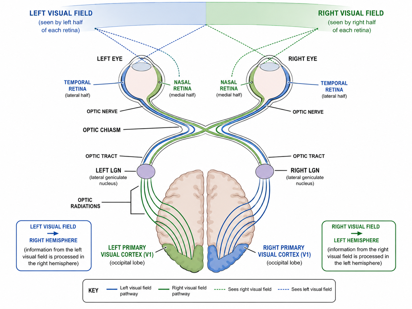
Not all retinal output goes to the cortex. A subset projects to the superior colliculus (midbrain), which controls reflexive eye movements (saccades) and orienting — shifting your gaze toward a sudden peripheral movement before you consciously decide to look.
| Lesion Location | Visual Deficit |
|---|---|
| Optic nerve (before chiasm) | Complete blindness in one eye (monocular blindness) |
| Optic chiasm (center) | Loss of both temporal (lateral) visual fields = bitemporal hemianopia |
| Optic tract / LGN / optic radiation | Loss of same side visual field in both eyes = homonymous hemianopia |
| Right V1 | Loss of left visual field in both eyes |
The classic cause of bitemporal hemianopia is a pituitary adenoma pressing on the chiasm from below — the crossing nasal fibers (carrying temporal visual field information) are selectively destroyed.
The LGN and Primary Visual Cortex
Must knowThe LGN of the thalamus is a relay that organizes inputs by eye and by cell type (magnocellular vs. parvocellular — see parallel processing).
V1 (primary visual cortex) in the occipital lobe is organized retinotopically — adjacent retinal points map to adjacent V1 points, with the fovea disproportionately represented (cortical magnification).
Quick check (scenario): A patient has a pituitary tumor and reports that they can only see objects directly in front of them but not to either side — they describe it as "looking through a tube." Which fibers were damaged, and where?
Answer: The crossing nasal retinal fibers at the optic chiasm were damaged. These fibers carry input from the temporal visual fields of each eye. Losing them on both sides produces bitemporal hemianopia.
Parallel Processing
Why Process Things in Parallel?
Must knowThe visual system analyzes form, color, motion, and depth simultaneously in specialized pathways. This is parallel processing — it's why you perceive a fast-moving red ball: motion and color are processed at once, then integrated.
Know the logicTwo streams originate in distinct RGCs:
- Magnocellular (M): large, fast cells → motion and depth → feeds the dorsal stream.
- Parvocellular (P): smaller, slower cells → color and fine detail → feeds the ventral stream.
The Two Cortical Streams
Must knowAfter V1, information splits into two routes:
- Dorsal stream ("where/how"): V1 → parietal lobe. Spatial location, motion, visually guided action. Damage → trouble reaching/navigating despite intact recognition.
- Ventral stream ("what"): V1 → temporal lobe. Object identity, form, color, face recognition. Damage → visual agnosia (can't recognize objects despite intact vision).
Mnemonic: dorsal = "where/how" (parietal, on top, like a map); ventral = "what" (temporal, where object memories live).
Quick check (scenario): A patient with a brain lesion can accurately reach out and grab a pen on a desk but cannot identify what the pen is just by looking at it. Which stream is likely damaged?
Answer: The ventral ("what") stream in the temporal lobe. The dorsal stream (spatial/action guidance) is intact — he can grab the object — but the ventral stream that identifies objects is impaired.
Feature Detection
The Neuroscience of "What Does This Cell See?"
Must knowIndividual cortical neurons respond to specific features of a stimulus (edges, orientations, movement). This is feature detection: specialized neurons fire maximally to particular visual properties.
Hubel and Wiesel — The Classic Experiment
Must knowKnow the name and the core finding. Hubel and Wiesel recorded from single neurons in cat V1 and found orientation-selective cells that extract features hierarchically:
- Simple cells: respond to edges at a specific orientation and location.
- Complex cells: respond to an oriented edge anywhere in a larger field, and to motion.
- Optional Hypercomplex (end-stopped) cells add length selectivity.
They also discovered critical periods: kittens deprived of patterned vision in one eye during a neonatal window became permanently blind in it — relevant to childhood disorders (e.g., uncorrected strabismus, amblyopia).
Gestalt Principles — How the Brain Groups Features
Know the logicPerception also requires organizing features into coherent objects. The Gestalt principles describe how we automatically group visual elements (recognize each by name):
| Gestalt Principle | Description |
|---|---|
| Figure–Ground | Separate a scene into figure (focus) vs. background (e.g., Rubin's vase). |
| Proximity | Close-together elements are grouped. |
| Similarity | Alike elements (color, shape) are grouped. |
| Continuity | Lines/curves perceived as continuing smoothly. |
| Closure | We fill in gaps to perceive a complete shape. |
| Prägnanz (Simplicity) | We perceive the simplest, most stable interpretation — the overarching law. |
| Common Fate | Elements moving the same direction are grouped. |
Depth Perception — Monocular and Binocular Cues
Know the logicPerceiving 3D from 2D retinal images uses two categories of cues.
Binocular cues (need both eyes):
- Retinal disparity: the two eyes' slightly different images; closer objects → greater disparity. Basis of stereopsis.
- Convergence: how far the eyes rotate inward to fixate; more convergence = closer.
Monocular cues: linear perspective, relative size, interposition (overlap), texture gradient, motion parallax (near things sweep by faster), aerial perspective (distant things hazier), and shading/shadow.
Perceptual Constancies
Know the logicPerceptual constancy — stable perception despite changing retinal images:
- Size constancy: true size perceived even as the retinal image shrinks with distance.
- Shape constancy: a door looks rectangular even when angled (trapezoid on retina).
- Color constancy: perceived color stays stable under different lighting.
Quick check (scenario): You are watching a flock of birds where each individual bird is too far away to identify, but the whole group appears to move together in the same direction, and you effortlessly perceive them as a unit. Which Gestalt principle is operating?
Answer: Common fate — elements moving in the same direction are grouped together. (Proximity also contributes since the birds are physically close.)
Common Confusions & Tricks
1. Rods depolarize in the dark. Dark is the resting state; light turns off the dark current (hyperpolarization). Not "activated by darkness."
2. Ciliary muscles contracted = near vision (not distance). Contraction releases tension on the zonular fibers, letting the lens round up. Think: "relax the string, the lens springs back to round."
3. Nasal fibers cross at the chiasm; temporal fibers don't. Your nose is in the middle — nasal fibers go to the middle of the chiasm and cross. Temporal (outer) fibers stay ipsilateral.
4. Bitemporal hemianopia = loss of temporal visual fields, not temporal retinal fibers. Nasal retinal fibers carry the temporal visual fields; the lesion cuts nasal fibers → loss of peripheral vision. The name is about what the patient can't see.
5. Trichromatic vs. opponent-process — both correct, at different levels. Three cone types = trichromatic; RGC/LGN opponent channels and afterimages = opponent-process.
6. Dorsal = "where/how," Ventral = "what." Temporal lobe (ventral) = what things are; parietal (dorsal) = where in space.
7. Hubel & Wiesel ≠ color vision. Their work = orientation/feature detection in V1. Color opponency = Hering; three-cone vision = Young–Helmholtz.
8. Presbyopia vs. hyperopia — both use converging lenses, different causes. Hyperopia = eyeball too short (structural); presbyopia = stiff lens (accommodation failure).
9. Afterimage question → opponent-process theory. Afterimage color = opponent of the original.
Key Theories & Terms
| Term / Name | What It Is / Who |
|---|---|
| Cornea | Transparent front surface of the eye; provides most of the eye's refractive power |
| Accommodation | Process by which the ciliary muscles contract, releasing zonular fiber tension and allowing the lens to round up for near-focus |
| Rods | Photoreceptors for dim (scotopic) vision; contain rhodopsin; peripheral retina; no color discrimination |
| Cones | Photoreceptors for bright (photopic) vision and color; three types (S, M, L); concentrated at the fovea |
| Fovea | Central region of the retina with highest cone density and highest visual acuity |
| Optic disc (blind spot) | Point where the optic nerve exits the retina; no photoreceptors; produces a blind spot in the visual field |
| Rhodopsin | Photopigment in rods, composed of opsin + 11-cis retinal; converts light to a neural signal |
| Dark current | Steady inward current that keeps photoreceptors depolarized in the dark; shut off by light |
| Phototransduction | Process converting light into a graded receptor potential that hyperpolarizes the photoreceptor and reduces glutamate release |
| Superior colliculus | Midbrain target of some retinal fibers; controls reflexive saccadic eye movements and orienting toward stimuli |
| Trichromatic theory (Young–Helmholtz) | Three cone types (S, M, L) underlie color vision; color is perceived by comparing relative activation ratios |
| Opponent-process theory (Hering) | Color and luminance are encoded in opponent pairs (red–green, blue–yellow, black–white) at the retinal/LGN level; explains afterimages |
| Myopia | Nearsightedness; image focuses in front of retina; corrected with diverging (concave) lens |
| Hyperopia | Farsightedness; image focuses behind retina; corrected with converging (convex) lens |
| Presbyopia | Age-related loss of accommodation due to lens rigidity; corrected with converging reading lenses |
| Optic chiasm | Point where nasal retinal fibers cross to the contralateral optic tract; each hemisphere receives contralateral visual field |
| Lateral Geniculate Nucleus (LGN) | Thalamic relay for visual information; organized in magnocellular and parvocellular layers |
| Primary visual cortex (V1, striate cortex) | Located in the occipital lobe; first cortical processing of visual input; retinotopic organization |
| Dorsal stream ("where/how" pathway) | V1 → parietal lobe; processes spatial location and visually guided action |
| Ventral stream ("what" pathway) | V1 → temporal lobe; processes object identity, color, and face recognition |
| Parallel processing | Simultaneous analysis of different visual attributes (motion, color, form, depth) by different neural pathways |
| Feature detection | The principle that specific cortical neurons respond maximally to specific visual features (edges, orientations, movement) |
| Hubel & Wiesel | Nobel laureates who discovered orientation-selective simple, complex, and hypercomplex cells in V1; also discovered visual critical periods |
| Simple cells | V1 neurons responding to edges at a specific orientation and location |
| Complex cells | V1 neurons responding to oriented edges anywhere in a larger receptive field; motion sensitive |
| Gestalt principles | A set of rules (proximity, similarity, continuity, closure, figure–ground, prägnanz, common fate) describing how the visual system automatically groups elements into organized percepts |
| Prägnanz | The overarching Gestalt principle: we perceive the simplest, most stable interpretation of visual scenes |
| Retinal disparity | The difference between the images of each eye used as a binocular depth cue; the basis of stereopsis |
| Size constancy | Perceptual stability of an object's perceived size despite changes in retinal image size with distance |
| Bitemporal hemianopia | Loss of both temporal visual fields due to a lesion of crossing nasal fibers at the optic chiasm (classic: pituitary adenoma) |
| Homonymous hemianopia | Loss of the same visual field in both eyes due to a lesion behind the optic chiasm (optic tract, LGN, radiation, or V1) |